Cookie preferences
This website uses cookies, which are necessary for the technical operation of the website and are always set. Other cookies, which increase the comfort when using this website, are used for direct advertising or to facilitate interaction with other websites and social networks, are only set with your consent.
Configuration
Technically required
These cookies are necessary for the basic functions of the shop.
"Allow all cookies" cookie
"Decline all cookies" cookie
CSRF token
Cookie preferences
Currency change
Customer-specific caching
FACT-Finder tracking
Individual prices
Selected shop
Session
Comfort functions
These cookies are used to make the shopping experience even more appealing, for example for the recognition of the visitor.
Note
Show the facebook fanpage in the right blod sidebar
Statistics & Tracking
Affiliate program
Conversion and usertracking via Google Tag Manager
Track device being used
| Item number | Size | Datasheet | Manual | SDS | Delivery time | Quantity | Price |
|---|---|---|---|---|---|---|---|
| NSJ-FY12572 | 100 µl | - | - |
3 - 10 business days* |
790.00€
|
If you have any questions, please use our Contact Form.
You can also order by e-mail: info@biomol.com
Larger quantity required? Request bulk
You can also order by e-mail: info@biomol.com
Larger quantity required? Request bulk
Rabbit IgG in phosphate buffered saline, pH 7.4, 150mM NaCl, 0.02% sodium azide and 50% glycerol,... more
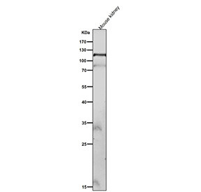
Product information "Anti-phospho-FAK (Tyr576/Tyr577) / Focal Adhesion Kinase 1, clone 32P46"
Rabbit IgG in phosphate buffered saline, pH 7.4, 150mM NaCl, 0.02% sodium azide and 50% glycerol, 0.4-0.5mg/ml BSA. Phospho-FAK (Tyr576/Tyr577) antibody detects focal adhesion kinase 1 when phosphorylated at tyrosine residues 576 and 577. Focal adhesion kinase, encoded by the PTK2 gene, is a cytoplasmic tyrosine kinase that regulates adhesion, migration, and survival signaling. Autophosphorylation at Tyr397 recruits Src family kinases, which subsequently phosphorylate Tyr576 and Tyr577 within the kinase activation loop. Phosphorylation at these residues enhances catalytic activity and promotes downstream signaling via MAPK and PI3K pathways.Phospho-FAK (Tyr576/Tyr577) antibody is widely used in cancer biology, cell adhesion studies, and mechanotransduction research. Active FAK drives tumor progression, metastasis, and angiogenesis by promoting migration and survival of cancer cells. Detection of phosphorylation at Tyr576/577 provides a direct measure of FAK activation and signaling output. By using this antibody, researchers can evaluate FAK driven pathways in oncogenesis, wound healing, and fibrosis. Western blot assays distinguish phosphorylated FAK from unmodified protein, while immunohistochemistry maps activation in tissues undergoing remodeling or tumor progression. Immunofluorescence highlights localization of phospho-FAK at focal adhesions, providing spatial insight into adhesion-dependent signaling events. These approaches allow comprehensive analysis of FAK activation in different systems.Phosphorylation at Tyr576 and Tyr577 is essential for full kinase activation. It promotes conformational changes that enhance substrate binding and signaling complex assembly. Dysregulated FAK phosphorylation contributes to cancer cell invasion and therapy resistance. By applying Phospho-FAK (Tyr576/Tyr577) antibody, scientists can study therapeutic strategies that target adhesion-dependent kinase activity and its role in disease.FAK signaling also participates in cardiovascular and developmental biology, where it regulates vascular remodeling, stem cell adhesion, and tissue morphogenesis. Its integration of mechanical and biochemical signals makes it a central regulator of cell behavior. The phospho-specific antibody therefore serves as a powerful tool for examining mechanosensitive signaling networks.Phospho-FAK (Tyr576/Tyr577) antibody from NSJ Bioreagents ensures accurate detection of activated PTK2 in cancer, adhesion, and mechanotransduction research.
| Keywords: | Anti-phospho-PTK2, Anti-phospho-p125FAK, Anti-phospho-pp125FAK, Anti-phospho-Focal adhesion kinase 1, Anti-phospho-Protein-tyrosine kinase 2, Anti-phospho-Focal adhesion kinase-related nonkinase, Anti-phospho-Protein phosphatase 1 regulatory subunit 71, P |
| Supplier: | NSJ Bioreagents |
| Supplier-Nr: | FY12572 |
Properties
| Application: | WB |
| Antibody Type: | Monoclonal |
| Clone: | 32P46 |
| Conjugate: | No |
| Host: | Rabbit |
| Species reactivity: | human, mouse |
| Immunogen: | A synthesized peptide derived from human Phospho-FAK (Y576 + Y577) |
| Format: | Purified |
Database Information
| KEGG ID : | K05725 | Matching products |
| UniProt ID : | Q05397 | Matching products |
| Gene ID : | GeneID 5747 | Matching products |
Handling & Safety
| Storage: | -20°C |
| Shipping: | -20°C (International: -20°C) |
Caution
Our products are for laboratory research use only: Not for administration to humans!
Our products are for laboratory research use only: Not for administration to humans!
Information about the product reference will follow.
more
You will get a certificate here
Viewed