Cookie preferences
This website uses cookies, which are necessary for the technical operation of the website and are always set. Other cookies, which increase the comfort when using this website, are used for direct advertising or to facilitate interaction with other websites and social networks, are only set with your consent.
Configuration
Technically required
These cookies are necessary for the basic functions of the shop.
"Allow all cookies" cookie
"Decline all cookies" cookie
CSRF token
Cookie preferences
Currency change
Customer-specific caching
FACT-Finder tracking
Individual prices
Selected shop
Session
Comfort functions
These cookies are used to make the shopping experience even more appealing, for example for the recognition of the visitor.
Note
Show the facebook fanpage in the right blod sidebar
Statistics & Tracking
Affiliate program
Conversion and usertracking via Google Tag Manager
Track device being used


| Item number | Size | Datasheet | Manual | SDS | Delivery time | Quantity | Price |
|---|---|---|---|---|---|---|---|
| NSJ-V6123-20UG | 20 µg | - | - |
3 - 10 business days* |
684.00€
|
||
| NSJ-V6123-100UG | 100 µg | - | - |
3 - 10 business days* |
1,372.00€
|
If you have any questions, please use our Contact Form.
You can also order by e-mail: info@biomol.com
Larger quantity required? Request bulk
You can also order by e-mail: info@biomol.com
Larger quantity required? Request bulk
Antibody in 1X PBS with 0.05% BSA, 0.05% sodium azide. DNA topoisomerase II alpha (TOP2A) is a... more
Product information "Anti-Topoisomerase II alpha / TOP2A, clone MSVA-802R"
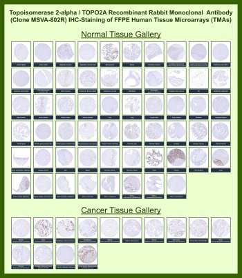
Antibody in 1X PBS with 0.05% BSA, 0.05% sodium azide. DNA topoisomerase II alpha (TOP2A) is a nuclear enzyme that regulates DNA topology during replication, transcription, and chromosome segregation. The protein belongs to the type II DNA topoisomerase family and functions by creating transient double strand breaks to relieve torsional stress during DNA replication. Because TOP2A expression is tightly linked to the S and G2/M phases of the cell cycle, it is widely recognized as a nuclear proliferation marker in rapidly dividing cells and malignant tumors.TOP2A Antibody for IHC / Topoisomerase II Alpha Immunohistochemistry Antibody (clone MSVA-802R) is designed specifically for immunohistochemistry based detection of TOP2A protein in formalin-fixed paraffin-embedded tissue sections. As a recombinant rabbit monoclonal antibody, clone MSVA-802R provides strong nuclear staining in proliferating cells, enabling clear visualization of TOP2A expression patterns within tumor tissue architecture. In immunohistochemistry analysis, TOP2A antibody staining typically appears as distinct nuclear labeling in mitotically active epithelial cells, germinal center lymphocytes, and rapidly proliferating tumor cells.Immunohistochemistry detection of TOP2A is widely used in cancer research and pathology because nuclear expression correlates with tumor proliferation rate. Many malignancies including breast carcinoma, lung carcinoma, colorectal carcinoma, and high grade lymphomas show strong nuclear staining when examined using a TOP2A antibody in immunohistochemistry assays. These staining patterns reflect the biological role of TOP2A as a cell cycle regulated enzyme required for chromosome condensation and segregation during mitosis. As a result, TOP2A antibody staining is commonly evaluated alongside other proliferation markers in tumor tissue studies.Evaluation of the TOP2A Antibody for IHC using human tissue microarray (TMA) panels provides an efficient method for examining nuclear staining patterns across a wide range of normal and cancer tissues. Tissue microarrays contain dozens to hundreds of tissue cores embedded in a single slide, allowing immunohistochemistry staining conditions to be applied uniformly across many tissue types. When clone MSVA-802R is applied to human tissue microarray slides, strong nuclear TOP2A immunoreactivity is typically observed in tumors with high proliferative activity, while most normal tissues display limited staining restricted to proliferative compartments.Tissue microarray immunohistochemistry analysis is particularly valuable for validating antibodies intended for pathology research. Because multiple tumor entities and normal tissues can be evaluated simultaneously, TMA based immunohistochemistry allows rapid comparison of nuclear staining intensity, cellular localization, and tumor specific expression patterns. The TOP2A Antibody for IHC therefore benefits from tissue microarray analysis where consistent nuclear staining can be assessed across large panels of human tissues under identical immunohistochemistry conditions.In immunohistochemistry studies, TOP2A antibody staining highlights nuclei of tumor cells undergoing active proliferation, making it a useful marker for studies focused on tumor growth and cell cycle activity. Nuclear localization detected with Topoisomerase II Alpha Immunohistochemistry Antibody reflects the enzyme's role in regulating DNA topology during chromosomal replication and segregation. Because TOP2A accumulates in dividing cells, immunohistochemistry staining intensity often parallels proliferative activity within tumor tissue sections.Use of tissue microarray slides further strengthens evaluation of TOP2A antibody performance in immunohistochemistry workflows. Large scale TMA screening allows visualization of nuclear TOP2A staining across diverse tumor types while maintaining consistent antigen retrieval and staining conditions. This approach provides valuable data for researchers examining proliferation related biomarkers in archival pathology specimens.Clone MSVA-802R enables robust nuclear detection of TOP2A in formalin-fixed paraffin-embedded tissues using immunohistochemistry. When applied to human tissue microarray panels containing multiple normal and malignant tissues, the antibody reveals characteristic nuclear staining patterns associated with proliferating cells. These features make the TOP2A Antibody for IHC a valuable tool for studies investigating tumor proliferation, DNA replication machinery, and cell cycle associated biomarkers in tissue-based immunohistochemistry analysis.This antibody is also part of a broader collection of IHC antibodies validated by tissue microarray analysis, supporting consistent staining across normal and cancer tissues.
| Keywords: | Anti-TOP2A, Anti-DNA topoisomerase 2-alpha, Anti-DNA topoisomerase II, alpha isozyme, Topoisomerase II alpha Antibody / TOP2A |
| Supplier: | NSJ Bioreagents |
| Supplier-Nr: | V6123 |
Properties
| Application: | IHC |
| Antibody Type: | Monoclonal |
| Clone: | MSVA-802R |
| Conjugate: | No |
| Host: | Rabbit |
| Species reactivity: | human |
| Immunogen: | Recombinant human Topoisomerase II alpha fragment (amino acids 1352-1493-exact sequence is proprietary) |
| Format: | Purified |
Database Information
| KEGG ID : | K03164 | Matching products |
| UniProt ID : | P11388 | Matching products |
| Gene ID : | GeneID 7153 | Matching products |
Handling & Safety
| Storage: | -20°C |
| Shipping: | -20°C (International: -20°C) |
Caution
Our products are for laboratory research use only: Not for administration to humans!
Our products are for laboratory research use only: Not for administration to humans!
Information about the product reference will follow.
more
You will get a certificate here
Viewed