Cookie preferences
This website uses cookies, which are necessary for the technical operation of the website and are always set. Other cookies, which increase the comfort when using this website, are used for direct advertising or to facilitate interaction with other websites and social networks, are only set with your consent.
Configuration
Technically required
These cookies are necessary for the basic functions of the shop.
"Allow all cookies" cookie
"Decline all cookies" cookie
CSRF token
Cookie preferences
Currency change
Customer-specific caching
FACT-Finder tracking
Individual prices
Selected shop
Session
Comfort functions
These cookies are used to make the shopping experience even more appealing, for example for the recognition of the visitor.
Note
Show the facebook fanpage in the right blod sidebar
Statistics & Tracking
Affiliate program
Conversion and usertracking via Google Tag Manager
Track device being used


| Item number | Size | Datasheet | Manual | SDS | Delivery time | Quantity | Price |
|---|---|---|---|---|---|---|---|
| NSJ-F49539-0.08ML | 80 µl | - | - |
3 - 10 business days* |
361.00€
|
||
| NSJ-F49539-0.4ML | 400 µl | - | - |
3 - 10 business days* |
772.00€
|
If you have any questions, please use our Contact Form.
You can also order by e-mail: info@biomol.com
Larger quantity required? Request bulk
You can also order by e-mail: info@biomol.com
Larger quantity required? Request bulk
In 1X PBS, pH 7.4, with 0.09% sodium azide. Tumor protein p53 (TP53) is a sequence-specific... more
Product information "Anti-p53 / TP53"
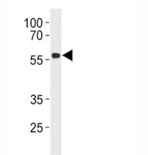
In 1X PBS, pH 7.4, with 0.09% sodium azide. Tumor protein p53 (TP53) is a sequence-specific transcription factor that functions as a central tumor suppressor regulating DNA damage responses, cell cycle arrest, apoptosis, and genomic stability. A TP53 Antibody for Immunofluorescence enables direct visualization of p53 localization within cells using fluorescence microscopy, allowing researchers to examine nuclear accumulation, intracellular distribution, and dynamic changes in p53 expression at the single-cell level.TP53 antibody, also referred to as Tumor protein p53 antibody or Cellular tumor antigen p53 antibody in the literature, targets one of the most extensively studied regulatory proteins in cancer biology. The TP53 gene is located on chromosome 17p13.1 and encodes a transcription factor belonging to the p53 family of DNA-binding proteins. The p53 protein contains several functional domains including an N-terminal transcriptional activation domain, a central DNA-binding region responsible for target gene recognition, a tetramerization domain that forms active p53 complexes, and a C-terminal regulatory region that modulates protein stability and DNA interaction.In unstressed cells, p53 protein levels remain extremely low due to continuous ubiquitination and proteasomal degradation mediated by the E3 ubiquitin ligase MDM2. When cells encounter stress signals such as DNA damage, oncogene activation, oxidative stress, or hypoxia, this degradation pathway is inhibited and p53 becomes stabilized. The stabilized protein rapidly accumulates in the nucleus where it activates transcription of genes including CDKN1A (p21), BAX, and PUMA. A TP53 Antibody for Immunofluorescence allows these stress-induced changes to be visualized through fluorescent nuclear staining that reflects activation of the p53 signaling pathway.Because many TP53 mutations produce stabilized proteins that accumulate in tumor cell nuclei, cancer cells frequently display strong nuclear fluorescence when stained using a TP53 Antibody for Immunofluorescence. Immunofluorescence imaging therefore provides a powerful method for detecting abnormal p53 accumulation and examining tumor-associated alterations in p53 regulation. In cancer cell lines, p53 staining commonly appears as bright nuclear fluorescence that contrasts with weak or absent signal in cells lacking stabilized protein.Immunofluorescence microscopy using a TP53 Antibody for Immunofluorescence is particularly valuable for studying the spatial organization of p53 within individual cells. Fluorescent imaging can reveal nuclear enrichment, redistribution following DNA damage, and cell-to-cell variability in p53 expression within heterogeneous populations. These imaging-based approaches allow researchers to monitor p53 pathway activation after chemotherapeutic treatment, radiation exposure, or experimental induction of cellular stress.Beyond its classical tumor suppressor function, p53 regulates numerous biological processes including metabolism, autophagy, immune signaling, and stem cell homeostasis. The protein interacts with multiple regulatory partners such as MDM2, ATM, ATR, and p300/CBP that influence transcriptional activity and intracellular localization. Because these regulatory interactions often alter nuclear abundance and spatial distribution of p53, fluorescence imaging using a TP53 Antibody for Immunofluorescence provides a valuable tool for studying p53 signaling dynamics and tumor suppressor activity in normal and transformed cells.
| Keywords: | Anti-TP53, Anti-Antigen NY-CO-13, Anti-Phosphoprotein p53, Anti-Tumor suppressor p53, Anti-Cellular tumor antigen p53, p53 Antibody / TP53 |
| Supplier: | NSJ Bioreagents |
| Supplier-Nr: | F49539 |
Properties
| Application: | IHC, IF, WB, ELISA |
| Antibody Type: | Polyclonal |
| Conjugate: | No |
| Host: | Rabbit |
| Species reactivity: | human |
| Immunogen: | Portion of amino acids 293-322 from the human protein |
| Format: | Purified |
Database Information
| KEGG ID : | K04451 | Matching products |
| UniProt ID : | P04637 | Matching products |
| Gene ID : | GeneID 7157 | Matching products |
Handling & Safety
| Storage: | -20°C |
| Shipping: | -20°C (International: -20°C) |
Caution
Our products are for laboratory research use only: Not for administration to humans!
Our products are for laboratory research use only: Not for administration to humans!
Information about the product reference will follow.
more
You will get a certificate here
Viewed