Cookie preferences
This website uses cookies, which are necessary for the technical operation of the website and are always set. Other cookies, which increase the comfort when using this website, are used for direct advertising or to facilitate interaction with other websites and social networks, are only set with your consent.
Configuration
Technically required
These cookies are necessary for the basic functions of the shop.
"Allow all cookies" cookie
"Decline all cookies" cookie
CSRF token
Cookie preferences
Currency change
Customer-specific caching
FACT-Finder tracking
Individual prices
Selected shop
Session
Comfort functions
These cookies are used to make the shopping experience even more appealing, for example for the recognition of the visitor.
Note
Show the facebook fanpage in the right blod sidebar
Statistics & Tracking
Affiliate program
Conversion and usertracking via Google Tag Manager
Track device being used


| Item number | Size | Datasheet | Manual | SDS | Delivery time | Quantity | Price |
|---|---|---|---|---|---|---|---|
| NSJ-V6124-20UG | 20 µg | - | - |
3 - 10 business days* |
684.00€
|
||
| NSJ-V6124-100UG | 100 µg | - | - |
3 - 10 business days* |
1,265.00€
|
If you have any questions, please use our Contact Form.
You can also order by e-mail: info@biomol.com
Larger quantity required? Request bulk
You can also order by e-mail: info@biomol.com
Larger quantity required? Request bulk
Antibody in 1X PBS with 0.05% BSA, 0.05% sodium azide. Tumor protein p53 (TP53) is a nuclear... more
Product information "Anti-p53 / TP53, clone MSVA-053R"
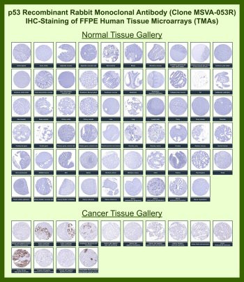
Antibody in 1X PBS with 0.05% BSA, 0.05% sodium azide. Tumor protein p53 (TP53) is a nuclear transcription factor that functions as one of the most extensively studied tumor suppressor proteins in human biology. Acting as a genomic surveillance protein, p53 regulates transcriptional programs that control cell cycle arrest, DNA repair, and apoptosis in response to cellular stress. The p53 Antibody for IHC / p53 Immunohistochemistry Antibody is designed for immunohistochemistry analysis of p53 protein expression in formalin-fixed, paraffin-embedded tissues, enabling visualization of nuclear p53 staining patterns in normal and tumor specimens.p53 antibody, also referred to as TP53 antibody or Tumor protein p53 antibody in the literature, is widely used for immunohistochemistry evaluation of p53 expression in pathological tissue samples. In healthy cells p53 protein levels remain low due to continuous ubiquitin-mediated degradation driven primarily by the E3 ubiquitin ligase MDM2. Cellular stress signals such as DNA damage, oncogene activation, oxidative stress, or hypoxia disrupt this regulatory pathway and allow accumulation of p53 in the nucleus, where it activates transcription of genes involved in growth arrest and programmed cell death. Because mutant forms of p53 often become stabilized in tumor cells, strong nuclear staining detected by immunohistochemistry frequently correlates with TP53 mutation status in many cancers.p53 immunohistochemistry has become a widely used method for studying tumor biology and evaluating p53 protein expression in FFPE tissues. Nuclear staining patterns observed with a p53 antibody for IHC reflect accumulation of p53 protein within tumor cells and are commonly examined in carcinomas of the colon, ovary, lung, breast, and many other tissues. Immunohistochemistry analysis allows pathologists and researchers to evaluate p53 expression within the architectural context of tissue sections, enabling assessment of tumor cell populations and their surrounding microenvironment.Tissue microarray (TMA) analysis provides an effective approach for evaluating antibody performance across a large panel of tissues simultaneously. Immunohistochemistry testing of recombinant rabbit monoclonal clone MSVA-053R using human tissue microarrays demonstrates nuclear staining patterns consistent with p53 expression in multiple tumor tissues while most normal tissues show limited staining. These TMA datasets enable side-by-side comparison of p53 immunohistochemistry staining across dozens of normal organs and cancer types, providing valuable reference information for evaluating p53 expression patterns in FFPE tissue specimens.Use of a p53 antibody for IHC allows researchers to visualize nuclear p53 protein distribution directly within tissue sections. Recombinant rabbit monoclonal antibody clone MSVA-053R is suitable for immunohistochemistry detection of p53 in FFPE tissues and supports tissue microarray-based studies investigating tumor suppressor biology, cancer pathology, and patterns of p53 expression across normal and malignant human tissues.This antibody is also part of a broader collection of IHC antibodies validated by tissue microarray analysis, supporting consistent staining across normal and cancer tissues.
| Keywords: | Anti-TP53, Anti-Antigen NY-CO-13, Anti-Phosphoprotein p53, Anti-Tumor suppressor p53, Anti-Cellular tumor antigen p53, p53 Antibody / TP53 |
| Supplier: | NSJ Bioreagents |
| Supplier-Nr: | V6124 |
Properties
| Application: | IHC |
| Antibody Type: | Monoclonal |
| Clone: | MSVA-053R |
| Conjugate: | No |
| Host: | Rabbit |
| Species reactivity: | human |
| Immunogen: | Recombinant human full-length TP53 protein |
| Format: | Purified |
Database Information
| KEGG ID : | K04451 | Matching products |
| UniProt ID : | P04637 | Matching products |
| Gene ID : | GeneID 7157 | Matching products |
Handling & Safety
| Storage: | -20°C |
| Shipping: | -20°C (International: -20°C) |
Caution
Our products are for laboratory research use only: Not for administration to humans!
Our products are for laboratory research use only: Not for administration to humans!
Information about the product reference will follow.
more
You will get a certificate here
Viewed