Cookie preferences
This website uses cookies, which are necessary for the technical operation of the website and are always set. Other cookies, which increase the comfort when using this website, are used for direct advertising or to facilitate interaction with other websites and social networks, are only set with your consent.
Configuration
Technically required
These cookies are necessary for the basic functions of the shop.
"Allow all cookies" cookie
"Decline all cookies" cookie
CSRF token
Cookie preferences
Currency change
Customer-specific caching
FACT-Finder tracking
Individual prices
Selected shop
Session
Comfort functions
These cookies are used to make the shopping experience even more appealing, for example for the recognition of the visitor.
Note
Show the facebook fanpage in the right blod sidebar
Statistics & Tracking
Affiliate program
Conversion and usertracking via Google Tag Manager
Track device being used
| Item number | Size | Datasheet | Manual | SDS | Delivery time | Quantity | Price |
|---|---|---|---|---|---|---|---|
| NSJ-FY13066 | 100 µg | - | - |
3 - 10 business days* |
790.00€
|
If you have any questions, please use our Contact Form.
You can also order by e-mail: info@biomol.com
Larger quantity required? Request bulk
You can also order by e-mail: info@biomol.com
Larger quantity required? Request bulk
Adding 0.2 ml of distilled water will yield a concentration of 500 ug/ml. MYL2 antibody detects... more
Product information "Anti-MYL2 / Myosin regulatory light chain 2"
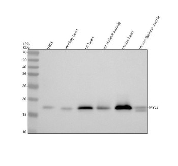
Adding 0.2 ml of distilled water will yield a concentration of 500 ug/ml. MYL2 antibody detects myosin regulatory light chain 2, a contractile protein critical for cardiac and skeletal muscle contraction. The UniProt recommended name is Myosin regulatory light chain 2 (MYL2). This 166-amino-acid protein binds to the neck region of myosin heavy chain, stabilizing the lever-arm structure and modulating actin-activated ATPase activity during force generation.Functionally, MYL2 antibody identifies a calcium-binding protein that undergoes phosphorylation by myosin light chain kinase (MLCK) at serine 15. Phosphorylation increases calcium sensitivity and enhances contractile force, playing a vital role in excitation-contraction coupling. MYL2 expression is strongest in cardiac ventricles, distinguishing it from atrial isoforms such as MYL7. Its presence marks ventricular myocytes in both mature and differentiating cardiac tissue.The MYL2 gene resides on chromosome 12q24.11 and encodes a regulatory component of the myosin complex. Mutations in MYL2 have been associated with familial hypertrophic cardiomyopathy, left ventricular dysfunction, and sudden cardiac death. These variants alter protein folding or phosphorylation sites, impairing actomyosin cross-bridge cycling and contractile mechanics.In muscle physiology, MYL2 acts as a fine-tuner of myosin motor function, adjusting contraction kinetics through dynamic phosphorylation. In developmental studies, MYL2 serves as a ventricular differentiation marker, while in pathology it reflects remodeling in cardiac hypertrophy and failure. Experimental models using MYL2 antibody provide insights into sarcomere assembly, cardiomyocyte maturation, and myofibrillar alignment.MYL2 antibody is suitable for western blotting, immunohistochemistry, and immunofluorescence to detect cardiac myosin light chain expression. It is extensively used in cardiovascular research, muscle biology, and regenerative medicine to monitor contractile protein regulation. NSJ Bioreagents offers validated MYL2 antibody reagents optimized for studies of cardiac physiology and disease.Structurally, MYL2 belongs to the EF-hand calcium-binding family and interacts with the IQ motif of myosin heavy chains. Its N-terminal domain modulates ATP hydrolysis and contributes to sarcomeric order. This antibody enables precise detection of MYL2 to study contractile regulation, disease mechanisms, and cardiomyocyte differentiation.
| Keywords: | Anti-MYL2, Anti-Cardiac myosin light chain 2, Anti-Ventricular myosin light chain 2, Anti-Myosin light chain 2, slow skeletal/ventricular muscle isoform, Anti-Myosin regulatory light chain 2, ventricular/cardiac muscle isoform, MYL2 Antibody / Myosin regu |
| Supplier: | NSJ Bioreagents |
| Supplier-Nr: | FY13066 |
Properties
| Application: | WB |
| Antibody Type: | Polyclonal |
| Conjugate: | No |
| Host: | Rabbit |
| Species reactivity: | human, monkey, mouse, rat |
| Immunogen: | A synthetic peptide corresponding to a sequence at the C-terminus of human Myosin Light Chain 2/MLC-2V/MYL2 |
| Format: | Purified |
Database Information
| KEGG ID : | K10351 | Matching products |
| UniProt ID : | P10916 | Matching products |
| Gene ID : | GeneID 4633 | Matching products |
Handling & Safety
| Storage: | +4°C |
| Shipping: | +4°C (International: +4°C) |
Caution
Our products are for laboratory research use only: Not for administration to humans!
Our products are for laboratory research use only: Not for administration to humans!
Information about the product reference will follow.
more
You will get a certificate here
Viewed