Cookie preferences
This website uses cookies, which are necessary for the technical operation of the website and are always set. Other cookies, which increase the comfort when using this website, are used for direct advertising or to facilitate interaction with other websites and social networks, are only set with your consent.
Configuration
Technically required
These cookies are necessary for the basic functions of the shop.
"Allow all cookies" cookie
"Decline all cookies" cookie
CSRF token
Cookie preferences
Currency change
Customer-specific caching
FACT-Finder tracking
Individual prices
Selected shop
Session
Comfort functions
These cookies are used to make the shopping experience even more appealing, for example for the recognition of the visitor.
Note
Show the facebook fanpage in the right blod sidebar
Statistics & Tracking
Affiliate program
Conversion and usertracking via Google Tag Manager
Track device being used


| Item number | Size | Datasheet | Manual | SDS | Delivery time | Quantity | Price |
|---|---|---|---|---|---|---|---|
| NSJ-V6155-20UG | 20 µg | - | - |
3 - 10 business days* |
917.00€
|
||
| NSJ-V6155-100UG | 100 µg | - | - |
3 - 10 business days* |
1,904.00€
|
If you have any questions, please use our Contact Form.
You can also order by e-mail: info@biomol.com
Larger quantity required? Request bulk
You can also order by e-mail: info@biomol.com
Larger quantity required? Request bulk
Antibody in 1X PBS with 0.05% BSA, 0.05% sodium azide. Methylthioadenosine phosphorylase (MTAP)... more
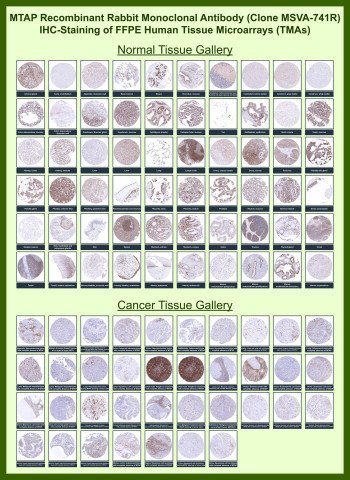
Product information "Anti-MTAP / S-methyl-5-thioadenosine phosphorylase, clone MSVA-741R"
Antibody in 1X PBS with 0.05% BSA, 0.05% sodium azide. Methylthioadenosine phosphorylase (MTAP) is a cytoplasmic metabolic enzyme that plays a central role in the methionine salvage pathway, catalyzing the conversion of methylthioadenosine to adenine and methylthioribose-1-phosphate. This reaction is essential for nucleotide recycling and maintenance of methionine homeostasis, linking MTAP activity directly to cellular metabolism and proliferation. MTAP (MTAP) is broadly expressed in normal tissues, particularly within epithelial and glandular compartments, where active metabolic turnover is required. MTAP Antibody for IHC / Tumor Metabolism Marker Antibody is optimized for detecting this enzyme in formalin-fixed, paraffin-embedded tissues, enabling evaluation of metabolic protein expression in histological context. For knockdown-validated detection of MTAP as a metabolic marker, see our MTAP antibody.MTAP antibody, also referred to as Methylthioadenosine phosphorylase antibody, demonstrates a characteristic cytoplasmic staining pattern in immunohistochemistry, consistent with the enzyme's intracellular localization. In normal human tissues, IHC analysis shows widespread and relatively uniform expression across a variety of organs, including epithelial linings, glandular structures, and metabolically active parenchymal cells. This baseline expression provides a clear reference for assessing changes in MTAP levels in disease states and supports its use in comparative tissue analysis.Tissue microarray (TMA) analysis across large panels of normal and cancer tissues highlights a defining feature of MTAP biology: its frequent loss in malignant cells. While normal tissues typically retain moderate to strong cytoplasmic staining, many tumor samples show markedly reduced or completely absent MTAP expression. This loss is observed across multiple tumor types and often appears as a sharp contrast between negative tumor regions and adjacent MTAP-positive stromal or non-neoplastic cells, creating a distinct and interpretable staining pattern. The inclusion of extensive TMA data strengthens confidence in the reproducibility of this expression profile across diverse tissue types and disease contexts.Functionally, MTAP loss is closely linked to alterations in tumor metabolism and cell cycle regulation. The MTAP gene is located near the CDKN2A locus on chromosome 9p21, and co-deletion of these regions is common in a wide range of cancers. As a result, loss of MTAP expression frequently accompanies disruption of tumor suppressor pathways, making it a useful surrogate marker in studies of genomic deletion events. In IHC, this manifests as absence of cytoplasmic staining in tumor cells despite preserved expression in surrounding normal tissue, providing a built-in internal control that enhances interpretability.In lymphoid tissues and other proliferative environments, MTAP staining may appear more heterogeneous, reflecting differences in metabolic activity among cell populations. In contrast, epithelial tumors often show more uniform loss or reduction of staining, depending on the extent of genomic alteration. These patterns are readily visualized in TMA formats, where parallel analysis of multiple tissue types enables direct comparison of staining intensity and distribution.Immunohistochemical staining with MTAP antibody clone MSVA-741R produces clear cytoplasmic signal with minimal background, supporting reliable identification of MTAP-positive and MTAP-negative cell populations. The combination of strong normal tissue expression, frequent tumor-associated loss, and robust performance in tissue microarrays makes this antibody particularly well suited for studies of tumor metabolism, biomarker discovery, and tissue-based analysis of gene deletion-associated phenotypes.This antibody is also part of a broader collection of IHC antibodies validated by tissue microarray analysis, supporting consistent staining across normal and cancer tissues.
| Keywords: | Anti-ERBB2, Anti-p185erbB2, Anti-Proto-oncogene Neu, Anti-Proto-oncogene c-ErbB-2, Anti-Metastatic lymph node gene 19 protein, Anti-Receptor tyrosine-protein kinase erbB-2, Anti-Tyrosine kinase-type cell surface receptor HER2, MTAP Antibody / S-methyl-5-t |
| Supplier: | NSJ Bioreagents |
| Supplier-Nr: | V6155 |
Properties
| Application: | IHC |
| Antibody Type: | Monoclonal |
| Clone: | MSVA-741R |
| Conjugate: | No |
| Host: | Rabbit |
| Species reactivity: | human |
| Immunogen: | Recombinant human MTAP protein fragment within amino acids 97-196 (exact sequence is proprietary) |
| Format: | Purified |
Database Information
| KEGG ID : | K05083 | Matching products |
| UniProt ID : | P04626 | Matching products |
| Gene ID : | GeneID 2064 | Matching products |
Handling & Safety
| Storage: | -20°C |
| Shipping: | -20°C (International: -20°C) |
Caution
Our products are for laboratory research use only: Not for administration to humans!
Our products are for laboratory research use only: Not for administration to humans!
Information about the product reference will follow.
more
You will get a certificate here
Viewed