Cookie preferences
This website uses cookies, which are necessary for the technical operation of the website and are always set. Other cookies, which increase the comfort when using this website, are used for direct advertising or to facilitate interaction with other websites and social networks, are only set with your consent.
Configuration
Technically required
These cookies are necessary for the basic functions of the shop.
"Allow all cookies" cookie
"Decline all cookies" cookie
CSRF token
Cookie preferences
Currency change
Customer-specific caching
FACT-Finder tracking
Individual prices
Selected shop
Session
Comfort functions
These cookies are used to make the shopping experience even more appealing, for example for the recognition of the visitor.
Note
Show the facebook fanpage in the right blod sidebar
Statistics & Tracking
Affiliate program
Conversion and usertracking via Google Tag Manager
Track device being used


| Item number | Size | Datasheet | Manual | SDS | Delivery time | Quantity | Price |
|---|---|---|---|---|---|---|---|
| NSJ-V6152-20UG | 20 µg | - | - |
3 - 10 business days* |
801.00€
|
||
| NSJ-V6152-100UG | 100 µg | - | - |
3 - 10 business days* |
1,436.00€
|
If you have any questions, please use our Contact Form.
You can also order by e-mail: info@biomol.com
Larger quantity required? Request bulk
You can also order by e-mail: info@biomol.com
Larger quantity required? Request bulk
Antibody in 1X PBS with 0.05% BSA, 0.05% sodium azide. Receptor tyrosine-protein kinase erbB-2... more
Product information "Anti-HER2 / ErbB2, clone MSVA-340R"
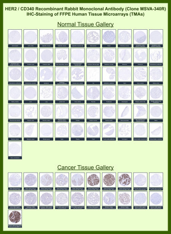
Antibody in 1X PBS with 0.05% BSA, 0.05% sodium azide. Receptor tyrosine-protein kinase erbB-2 (ERBB2), commonly known as HER2, is a transmembrane receptor tyrosine kinase of the EGFR family that plays a central role in epithelial cell signaling, proliferation, and survival. HER2 Antibody for IHC is widely used to assess HER2 protein expression in formalin-fixed, paraffin-embedded tissues, where its membranous localization and expression intensity provide critical information for tumor classification. In immunohistochemistry, HER2 staining is characterized by distinct membranous patterns that reflect receptor overexpression, making it one of the most clinically relevant epithelial biomarkers in pathology. In normal tissues, HER2 expression is typically low, with minimal or weak membranous staining confined to select epithelial compartments. This antibody is also part of a broader collection of IHC antibodies validated by tissue microarray analysis, supporting consistent staining across normal and cancer tissues.HER2 antibody, also referred to as ERBB2 antibody or HER2/neu antibody in the literature, recognizes a receptor that is frequently overexpressed in epithelial malignancies. This HER2 Antibody for IHC is specifically optimized for Tissue Microarray (TMA)-based immunohistochemistry, enabling high-throughput evaluation of HER2 expression across large panels of normal and cancer tissues. In normal tissue TMAs, staining is largely absent or weak, providing a clean background that enhances contrast when evaluating tumor samples. In cancer tissue microarrays, strong and specific membranous HRP-DAB brown staining is observed in HER2-positive tumors, particularly in breast carcinoma, where HER2 overexpression is a defining molecular feature.Tissue Microarray (TMA) analysis highlights the characteristic staining patterns of HER2 in tumor samples, including strong, circumferential membrane staining in HER2-positive breast cancers and variable membranous expression in gastric and other epithelial malignancies. HER2 Antibody for IHC enables clear differentiation between HER2-positive and HER2-negative tumors, supporting stratification of tumor subtypes based on receptor expression. The ability to assess staining intensity and membrane completeness across hundreds of cores within a single TMA slide allows consistent comparison of expression patterns, making this approach highly valuable for large-scale immunohistochemistry studies.The use of TMA-based IHC further demonstrates the specificity of clone MSVA-340R, with strong tumor-associated membranous staining observed alongside minimal background in non-epithelial and stromal tissues. This contrast enhances interpretability and supports accurate identification of HER2-expressing tumor cells within complex tissue environments. The reproducibility of staining across diverse TMA panels aligns with established HER2 expression data, including profiles reported in the Human Protein Atlas, reinforcing confidence in the antibody's performance in FFPE tissue analysis.This antibody targets HER2 in research applications requiring precise immunohistochemical detection of membranous receptor expression in FFPE tissue sections, making it well suited for studies of tumor classification, epithelial differentiation, and receptor-driven signaling in cancer biology. For broad detection of HER2 (ErbB2) as a receptor tyrosine kinase, see our HER2 antibody.
| Keywords: | Anti-ERBB2, Anti-p185erbB2, Anti-Proto-oncogene Neu, Anti-Proto-oncogene c-ErbB-2, Anti-Metastatic lymph node gene 19 protein, Anti-Receptor tyrosine-protein kinase erbB-2, Anti-Tyrosine kinase-type cell surface receptor HER2, HER2 Antibody / ErbB2 |
| Supplier: | NSJ Bioreagents |
| Supplier-Nr: | V6152 |
Properties
| Application: | IHC |
| Antibody Type: | Monoclonal |
| Clone: | MSVA-340R |
| Conjugate: | No |
| Host: | Rabbit |
| Species reactivity: | human |
| Immunogen: | Recombinant protein encoding the extracellular domain of human c-erbB2 |
| Format: | Purified |
Database Information
| KEGG ID : | K05083 | Matching products |
| UniProt ID : | P04626 | Matching products |
| Gene ID : | GeneID 2064 | Matching products |
Handling & Safety
| Storage: | -20°C |
| Shipping: | -20°C (International: -20°C) |
Caution
Our products are for laboratory research use only: Not for administration to humans!
Our products are for laboratory research use only: Not for administration to humans!
Information about the product reference will follow.
more
You will get a certificate here
Viewed