
Cookie preferences
This website uses cookies, which are necessary for the technical operation of the website and are always set. Other cookies, which increase the comfort when using this website, are used for direct advertising or to facilitate interaction with other websites and social networks, are only set with your consent.
Configuration
Technically required
These cookies are necessary for the basic functions of the shop.
"Allow all cookies" cookie
"Decline all cookies" cookie
CSRF token
Cookie preferences
Currency change
Customer-specific caching
FACT-Finder tracking
Individual prices
Selected shop
Session
Comfort functions
These cookies are used to make the shopping experience even more appealing, for example for the recognition of the visitor.
Note
Show the facebook fanpage in the right blod sidebar
Statistics & Tracking
Affiliate program
Conversion and usertracking via Google Tag Manager
Track device being used








| Item number | Size | Datasheet | Manual | SDS | Delivery time | Quantity | Price |
|---|---|---|---|---|---|---|---|
| NSJ-F48161-0.08ML | 80 µl | - | - |
3 - 10 business days* |
361.00€
|
||
| NSJ-F48161-0.4ML | 400 µl | - | - |
3 - 10 business days* |
772.00€
|
If you have any questions, please use our Contact Form.
You can also order by e-mail: info@biomol.com
Larger quantity required? Request bulk
You can also order by e-mail: info@biomol.com
Larger quantity required? Request bulk
In 1X PBS, pH 7.4, with 0.09% sodium azide. Vimentin (VIM) is a type III intermediate filament... more
Product information "Anti-Vimentin"
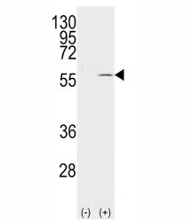
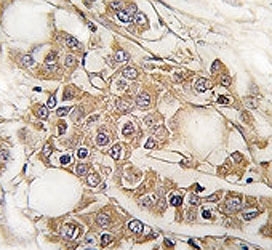
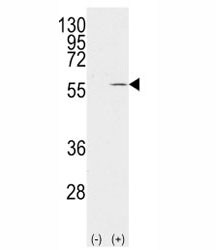
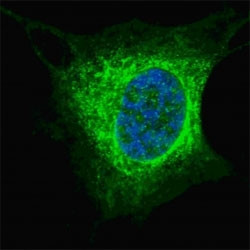
In 1X PBS, pH 7.4, with 0.09% sodium azide. Vimentin (VIM) is a type III intermediate filament protein that forms a highly organized cytoplasmic filament network in mesenchymal cells, where it maintains structural integrity and regulates cellular morphology. Vimentin Antibody for IF / Cytoskeletal Filament Marker Antibody is specifically optimized for immunofluorescence-based visualization of Vimentin filament architecture, enabling high-resolution imaging of cytoskeletal networks in fixed and permeabilized cells. This aligns strongly with search intent such as Vimentin immunofluorescence antibody, VIM IF antibody, and cytoskeletal filament marker antibody. Vimentin is also referred to as Vimentin antibody, VIM antibody, and intermediate filament marker antibody in the literature, supporting strong alignment between gene, protein, and imaging-focused terminology.In immunofluorescence and confocal microscopy applications, Vimentin forms a distinctive filamentous network that extends from the perinuclear region outward to the cell periphery. The Vimentin Antibody for IF / Cytoskeletal Filament Marker Antibody highlights these elongated cytoplasmic fibers, allowing researchers to directly visualize filament organization, network density, and structural remodeling within individual cells. This filament-specific staining pattern is a defining feature of the cytoskeletal filament marker antibody positioning and is essential for studies examining cytoskeletal architecture, cell polarity, and mechanical stability.Vimentin is widely expressed in fibroblasts, endothelial cells, leukocytes, and other mesenchymal-derived populations, where its filament network supports migration, adhesion, and intracellular transport. In fluorescence imaging workflows, the Vimentin Antibody for IF / Cytoskeletal Filament Marker Antibody enables detailed assessment of cytoskeletal rearrangement during processes such as cell spreading, migration, and epithelial-mesenchymal transition. It is also commonly used in co-localization studies alongside actin, tubulin, or organelle markers to evaluate spatial relationships between cytoskeletal systems.Functionally, Vimentin interacts with actin filaments and microtubules to coordinate cytoskeletal dynamics and intracellular organization. Its assembly and disassembly are regulated through phosphorylation and other post-translational modifications, which drive filament reorganization during mitosis and stress responses. The Vimentin Antibody for IF / Cytoskeletal Filament Marker Antibody differentiator is central to this antibody, positioning it specifically for fluorescence imaging applications where visualization of filament structure, network continuity, and subcellular distribution is critical. A rabbit polyclonal Vimentin Antibody for IF provides strong fluorescent signal and clear filament resolution, supporting high-quality imaging of Vimentin / VIM cytoskeletal networks in single-cell and multi-cellular systems.
| Keywords: | Anti-VIM, Anti-Vimentin, Vimentin Antibody |
| Supplier: | NSJ Bioreagents |
| Supplier-Nr: | F48161 |
Properties
| Application: | IF, WB, IHC, ELISA |
| Antibody Type: | Polyclonal |
| Conjugate: | No |
| Host: | Rabbit |
| Species reactivity: | human, mouse |
| Immunogen: | Portion of amino acids 63-90 from the human protein |
| Format: | Purified |
Database Information
| KEGG ID : | K07606 | Matching products |
| UniProt ID : | P08670 | Matching products |
| Gene ID : | GeneID 7431 | Matching products |
Handling & Safety
| Storage: | -20°C |
| Shipping: | -20°C (International: -20°C) |
Caution
Our products are for laboratory research use only: Not for administration to humans!
Our products are for laboratory research use only: Not for administration to humans!
Information about the product reference will follow.
more
You will get a certificate here
Viewed