
Cookie preferences
This website uses cookies, which are necessary for the technical operation of the website and are always set. Other cookies, which increase the comfort when using this website, are used for direct advertising or to facilitate interaction with other websites and social networks, are only set with your consent.
Configuration
Technically required
These cookies are necessary for the basic functions of the shop.
"Allow all cookies" cookie
"Decline all cookies" cookie
CSRF token
Cookie preferences
Currency change
Customer-specific caching
FACT-Finder tracking
Individual prices
Selected shop
Session
Comfort functions
These cookies are used to make the shopping experience even more appealing, for example for the recognition of the visitor.
Note
Show the facebook fanpage in the right blod sidebar
Statistics & Tracking
Affiliate program
Conversion and usertracking via Google Tag Manager
Track device being used


| Item number | Size | Datasheet | Manual | SDS | Delivery time | Quantity | Price |
|---|---|---|---|---|---|---|---|
| NSJ-V6128-20UG | 20 µg | - | - |
7 - 12 business days* |
684.00€
|
||
| NSJ-V6128-100UG | 100 µg | - | - |
7 - 12 business days* |
1,160.00€
|
If you have any questions, please use our Contact Form.
You can also order by e-mail: info@biomol.com
Larger quantity required? Request bulk
You can also order by e-mail: info@biomol.com
Larger quantity required? Request bulk
Antibody in 1X PBS with 0.05% BSA, 0.05% sodium azide. Villin-1 (VIL1) is an actin-associated... more
Product information "Anti-Villin, clone MSVA-459R"
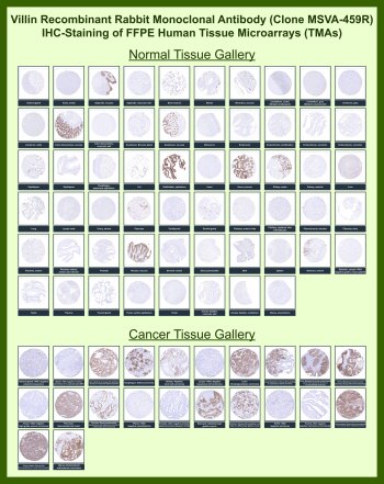
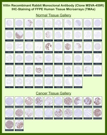
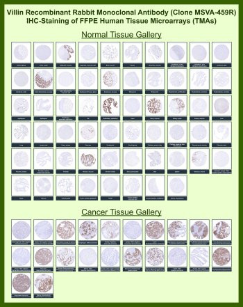
Antibody in 1X PBS with 0.05% BSA, 0.05% sodium azide. Villin-1 (VIL1) is an actin-associated protein localized to the apical region of intestinal epithelial cells, where it supports brush border organization and epithelial architecture. Villin Antibody for IHC / VIL1 Immunohistochemistry Antibody (clone MSVA-459R) is specifically developed for immunohistochemistry applications, enabling consistent detection of Villin in formalin-fixed, paraffin-embedded (FFPE) tissue sections. Villin antibody, also known as Villin-1 antibody or VIL1 antibody, is widely used in histological analysis to visualize epithelial structures and glandular morphology in gastrointestinal tissues.This Villin Antibody for IHC is strongly differentiated by its focus on immunohistochemistry performance, FFPE compatibility, and large-scale tissue validation rather than purely biological function. The recombinant rabbit monoclonal format (clone MSVA-459R) supports high signal clarity and reproducibility, which is critical for IHC workflows. Importantly, this antibody has been evaluated using human tissue microarray (TMA) panels, demonstrating consistent staining across a broad range of normal and cancer tissues. This TMA-based validation provides confidence in staining specificity and pattern consistency in complex tissue environments.In immunohistochemistry, Villin produces a characteristic apical and cytoplasmic staining pattern in epithelial cells, clearly outlining luminal borders and glandular structures. This makes Villin Antibody for IHC particularly valuable for assessing epithelial organization, identifying gland-forming tissues, and visualizing enterocyte-like differentiation in tumors. In colorectal and other gastrointestinal adenocarcinomas, Villin staining highlights tumor epithelial cells and supports evaluation of epithelial morphology within FFPE sections.From a pathology and research perspective, this VIL1 Immunohistochemistry Antibody is optimized for tissue-based analysis, including FFPE sections and TMA studies, where reproducible staining and clear morphological definition are essential. Clone MSVA-459R provides a robust tool for investigators and pathologists studying epithelial architecture, tumor histology, and gastrointestinal tissue identity using immunohistochemistry.This antibody is also part of a broader collection of IHC antibodies validated by tissue microarray analysis, supporting consistent staining across normal and cancer tissues.
| Keywords: | Anti-VIL1, Anti-Villin-1, Villin Antibody |
| Supplier: | NSJ Bioreagents |
| Supplier-Nr: | V6128 |
Properties
| Application: | IHC |
| Antibody Type: | Monoclonal |
| Clone: | MSVA-459R |
| Conjugate: | No |
| Host: | Rabbit |
| Species reactivity: | human |
| Immunogen: | Recombinant fragment (around amino acids 600-700) of human Villin protein (exact sequence is proprietary) |
| Format: | Purified |
Database Information
| KEGG ID : | K05761 | Matching products |
| UniProt ID : | P09327 | Matching products |
| Gene ID : | GeneID 7429 | Matching products |
Handling & Safety
| Storage: | -20°C |
| Shipping: | -20°C (International: -20°C) |
Caution
Our products are for laboratory research use only: Not for administration to humans!
Our products are for laboratory research use only: Not for administration to humans!
Information about the product reference will follow.
more
You will get a certificate here
Viewed