Cookie preferences
This website uses cookies, which are necessary for the technical operation of the website and are always set. Other cookies, which increase the comfort when using this website, are used for direct advertising or to facilitate interaction with other websites and social networks, are only set with your consent.
Configuration
Technically required
These cookies are necessary for the basic functions of the shop.
"Allow all cookies" cookie
"Decline all cookies" cookie
CSRF token
Cookie preferences
Currency change
Customer-specific caching
FACT-Finder tracking
Individual prices
Selected shop
Session
Comfort functions
These cookies are used to make the shopping experience even more appealing, for example for the recognition of the visitor.
Note
Show the facebook fanpage in the right blod sidebar
Statistics & Tracking
Affiliate program
Conversion and usertracking via Google Tag Manager
Track device being used


| Item number | Size | Datasheet | Manual | SDS | Delivery time | Quantity | Price |
|---|---|---|---|---|---|---|---|
| NSJ-V6117-20UG | 20 µg | - | - |
3 - 10 business days* |
684.00€
|
||
| NSJ-V6117-100UG | 100 µg | - | - |
3 - 10 business days* |
1,265.00€
|
If you have any questions, please use our Contact Form.
You can also order by e-mail: info@biomol.com
Larger quantity required? Request bulk
You can also order by e-mail: info@biomol.com
Larger quantity required? Request bulk
Antibody in 1X PBS with 0.05% BSA, 0.05% sodium azide. Transcription factor E3 (TFE3) is a member... more
Product information "Anti-TFE3 / Transcription factor E3, clone MSVA-403R"
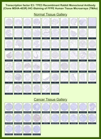
Antibody in 1X PBS with 0.05% BSA, 0.05% sodium azide. Transcription factor E3 (TFE3) is a member of the MiT family of basic helix-loop-helix leucine zipper transcription factors that regulate genes involved in cellular metabolism, lysosomal biogenesis, and autophagy. TFE3 Antibody for IHC / Xp11 Translocation Tumor Marker (clone MSVA-403R) is designed for immunohistochemistry analysis of tissue sections where detection of TFE3 protein provides a valuable nuclear biomarker for tumor evaluation. Immunohistochemistry staining allows pathologists to visualize TFE3 expression directly within preserved tissue architecture, enabling identification of tumor cells that display characteristic nuclear TFE3 staining patterns.TFE3 functions as a nuclear transcription factor that binds DNA regulatory elements to control gene expression programs involved in cellular growth and metabolic adaptation. Because the protein normally localizes to the cell nucleus, immunohistochemistry staining using a TFE3 antibody produces nuclear TFE3 staining in positive cells. This nuclear staining pattern is a key feature evaluated in diagnostic pathology when examining tissue sections for tumors associated with TFE3 gene rearrangements.Chromosomal translocations involving the Xp11 locus can lead to TFE3 gene fusions that cause abnormal overexpression of the TFE3 protein. These alterations are characteristic of Xp11 translocation tumors, including Xp11 translocation renal cell carcinoma and other neoplasms. In these tumors, immunohistochemistry detection of TFE3 frequently reveals strong nuclear TFE3 staining within tumor cells, providing an important morphological indicator that supports tumor classification during pathological examination.Clone MSVA-403R is a recombinant rabbit monoclonal antibody developed to detect TFE3 protein in formalin-fixed paraffin-embedded tissue sections. Recombinant rabbit monoclonal antibodies provide highly consistent target recognition and strong signal detection in tissue-based assays, making them well suited for immunohistochemistry applications where clear nuclear staining patterns must be interpreted within complex tissue environments.Tissue microarray (TMA) analysis represents a powerful validation approach for immunohistochemistry antibodies because it allows simultaneous evaluation of staining patterns across large panels of normal and tumor tissues. Immunohistochemistry testing of TMA sections using a TFE3 antibody enables researchers and pathologists to compare nuclear TFE3 staining across multiple tumor types under identical staining conditions. This approach helps confirm antibody specificity and provides valuable insight into the distribution of TFE3 expression in diverse tissues and malignancies.Because TFE3 functions as a nuclear transcription factor and a biomarker of Xp11 translocation tumors, immunohistochemistry detection of nuclear TFE3 staining remains an important tool for both research and diagnostic pathology. Detection of TFE3 using clone MSVA-403R supports studies of transcription factor activity, tumor biology, and evaluation of nuclear TFE3 staining patterns across tissue microarray panels and individual tumor specimens.This antibody is also part of a broader collection of IHC antibodies validated by tissue microarray analysis, supporting consistent staining across normal and cancer tissues.
| Keywords: | Anti-TFE3, Anti-Transcription factor E3, Anti-Class E basic helix-loop-helix protein 33, TFE3 Antibody / Transcription factor E3 |
| Supplier: | NSJ Bioreagents |
| Supplier-Nr: | V6117 |
Properties
| Application: | IHC |
| Antibody Type: | Monoclonal |
| Clone: | MSVA-403R |
| Conjugate: | No |
| Host: | Rabbit |
| Species reactivity: | human |
| Immunogen: | Recombinant full-length human TFE3 protein |
| Format: | Purified |
Database Information
| KEGG ID : | K09105 | Matching products |
| UniProt ID : | P19532 | Matching products |
| Gene ID : | GeneID 7030 | Matching products |
Handling & Safety
| Storage: | -20°C |
| Shipping: | -20°C (International: -20°C) |
Caution
Our products are for laboratory research use only: Not for administration to humans!
Our products are for laboratory research use only: Not for administration to humans!
Information about the product reference will follow.
more
You will get a certificate here
Viewed