Cookie preferences
This website uses cookies, which are necessary for the technical operation of the website and are always set. Other cookies, which increase the comfort when using this website, are used for direct advertising or to facilitate interaction with other websites and social networks, are only set with your consent.
Configuration
Technically required
These cookies are necessary for the basic functions of the shop.
"Allow all cookies" cookie
"Decline all cookies" cookie
CSRF token
Cookie preferences
Currency change
Customer-specific caching
FACT-Finder tracking
Individual prices
Selected shop
Session
Comfort functions
These cookies are used to make the shopping experience even more appealing, for example for the recognition of the visitor.
Note
Show the facebook fanpage in the right blod sidebar
Statistics & Tracking
Affiliate program
Conversion and usertracking via Google Tag Manager
Track device being used


| Item number | Size | Datasheet | Manual | SDS | Delivery time | Quantity | Price |
|---|---|---|---|---|---|---|---|
| NSJ-V6116-20UG | 20 µg | - | - |
3 - 10 business days* |
684.00€
|
||
| NSJ-V6116-100UG | 100 µg | - | - |
3 - 10 business days* |
1,160.00€
|
If you have any questions, please use our Contact Form.
You can also order by e-mail: info@biomol.com
Larger quantity required? Request bulk
You can also order by e-mail: info@biomol.com
Larger quantity required? Request bulk
Antibody in 1X PBS with 0.05% BSA, 0.05% sodium azide. Synaptophysin (SYP) is a synaptic vesicle... more
Product information "Anti-Synaptophysin, clone MSVA-462R"
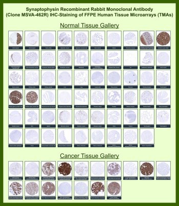
Antibody in 1X PBS with 0.05% BSA, 0.05% sodium azide. Synaptophysin (SYP) is a synaptic vesicle glycoprotein widely expressed in neurons and neuroendocrine cells where it functions as a major component of synaptic vesicle membranes involved in neurotransmitter storage and vesicle trafficking. Synaptophysin Antibody for IHC / SYP Immunohistochemistry Antibody (clone MSVA-462R) is a recombinant rabbit monoclonal antibody developed specifically for immunohistochemistry staining of Synaptophysin protein in formalin-fixed paraffin-embedded (FFPE) tissue sections. Synaptophysin is one of the most widely used neuroendocrine markers in immunohistochemistry and is routinely used in pathology to identify tumors with neuroendocrine differentiation. Because of its robust and characteristic staining pattern, Synaptophysin immunohistochemistry is commonly used to evaluate neuroendocrine tumors including small cell carcinoma, carcinoid tumors, pancreatic neuroendocrine tumors, neuroblastoma, and other malignancies demonstrating neuroendocrine lineage differentiation.In immunohistochemistry staining of FFPE tissues, Synaptophysin typically shows a distinctive granular cytoplasmic staining pattern corresponding to synaptic vesicle membranes within neuronal and neuroendocrine cells. Synaptophysin Antibody for IHC enables visualization of these vesicle-associated structures within tissue sections, allowing identification of neuroendocrine cell populations in both normal tissues and tumors. Immunohistochemistry detection of SYP therefore provides an important tool for studying neuroendocrine differentiation and for evaluating tumor samples where neuroendocrine lineage markers are being investigated. The characteristic punctate cytoplasmic staining observed by immunohistochemistry reflects the localization of Synaptophysin within synaptic vesicle membranes.Clone MSVA-462R is produced as a recombinant rabbit monoclonal antibody, combining the high affinity epitope recognition typical of rabbit-derived antibodies with the consistency of recombinant antibody technology. Recombinant rabbit monoclonal antibodies are generated from defined antibody sequences expressed in controlled recombinant systems, allowing highly reproducible antibody production and consistent immunohistochemistry staining results across manufacturing batches. The recombinant rabbit monoclonal format supports reliable detection of Synaptophysin protein in FFPE tissues and provides consistent staining performance for immunohistochemistry experiments examining neuroendocrine markers.Extensive validation using human tissue microarray (TMA) panels further supports the immunohistochemistry performance of this antibody. Tissue microarrays contain hundreds of individual tissue cores representing diverse normal organs and multiple tumor types, enabling systematic immunohistochemistry evaluation of protein expression patterns across large tissue cohorts. TMA-based immunohistochemistry analysis with Synaptophysin Antibody for IHC demonstrates cytoplasmic staining patterns consistent with known Synaptophysin expression in neuronal and neuroendocrine tissues while allowing comparison of expression across numerous tumor types. Human tissue microarray validation provides important evidence supporting antibody performance in immunohistochemistry by enabling evaluation of staining specificity, tissue distribution, and tumor-associated expression patterns across extensive normal and cancer tissue panels.Synaptophysin Antibody for IHC / SYP Immunohistochemistry Antibody (clone MSVA-462R) therefore provides a recombinant rabbit monoclonal antibody optimized for immunohistochemistry detection of the Synaptophysin synaptic vesicle protein. The combination of recombinant rabbit monoclonal antibody technology and large-scale human tissue microarray immunohistochemistry analysis supports reliable visualization of SYP expression in studies investigating neuroendocrine differentiation, neuronal biology, and neuroendocrine tumor pathology.This antibody is also part of a broader collection of IHC antibodies validated by tissue microarray analysis, supporting consistent staining across normal and cancer tissues.
| Keywords: | Anti-SYP, Anti-Synaptophysin, Anti-Major synaptic vesicle protein p38, Synaptophysin Antibody |
| Supplier: | NSJ Bioreagents |
| Supplier-Nr: | V6116 |
Properties
| Application: | IHC |
| Antibody Type: | Monoclonal |
| Clone: | MSVA-462R |
| Conjugate: | No |
| Host: | Rabbit |
| Species reactivity: | human |
| Immunogen: | Recombinant fragment (around amino acids 274-313) of human Synaptophysin (SYP) protein (exact sequence is proprietary) |
| Format: | Purified |
Database Information
| KEGG ID : | K28145 | Matching products |
| UniProt ID : | P08247 | Matching products |
| Gene ID : | GeneID 6855 | Matching products |
Handling & Safety
| Storage: | -20°C |
| Shipping: | -20°C (International: -20°C) |
Caution
Our products are for laboratory research use only: Not for administration to humans!
Our products are for laboratory research use only: Not for administration to humans!
Information about the product reference will follow.
more
You will get a certificate here
Viewed