Cookie preferences
This website uses cookies, which are necessary for the technical operation of the website and are always set. Other cookies, which increase the comfort when using this website, are used for direct advertising or to facilitate interaction with other websites and social networks, are only set with your consent.
Configuration
Technically required
These cookies are necessary for the basic functions of the shop.
"Allow all cookies" cookie
"Decline all cookies" cookie
CSRF token
Cookie preferences
Currency change
Customer-specific caching
FACT-Finder tracking
Individual prices
Selected shop
Session
Comfort functions
These cookies are used to make the shopping experience even more appealing, for example for the recognition of the visitor.
Note
Show the facebook fanpage in the right blod sidebar
Statistics & Tracking
Affiliate program
Conversion and usertracking via Google Tag Manager
Track device being used


| Item number | Size | Datasheet | Manual | SDS | Delivery time | Quantity | Price |
|---|---|---|---|---|---|---|---|
| NSJ-V6111-20UG | 20 µg | - | - |
3 - 10 business days* |
684.00€
|
||
| NSJ-V6111-100UG | 100 µg | - | - |
3 - 10 business days* |
1,160.00€
|
If you have any questions, please use our Contact Form.
You can also order by e-mail: info@biomol.com
Larger quantity required? Request bulk
You can also order by e-mail: info@biomol.com
Larger quantity required? Request bulk
Antibody in 1X PBS with 0.05% BSA, 0.05% sodium azide. Premelanosome protein (PMEL) is a... more
Product information "Anti-PMEL17 / Melanoma gp100, clone MSVA-617R"
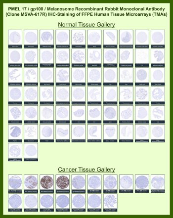
Antibody in 1X PBS with 0.05% BSA, 0.05% sodium azide. Premelanosome protein (PMEL) is a melanocyte lineage-associated glycoprotein encoded by the PMEL gene and widely known in the literature as gp100 or Pmel17. This melanosome-associated protein is expressed in melanocytes and melanocytic tumors and serves as a well-established melanocyte lineage marker in dermatopathology. gp100 Antibody for IHC / PMEL Melanocyte Lineage Marker (clone MSVA-617R) is designed for immunohistochemistry detection of melanocytic cells in formalin-fixed paraffin-embedded tissue sections. In immunohistochemistry studies, gp100 staining highlights melanocytes and melanoma tumor cells, supporting evaluation of melanocytic differentiation in tissue specimens.Immunohistochemistry analysis of PMEL typically demonstrates cytoplasmic staining within melanocytes located along the basal layer of the epidermis in normal skin. In melanoma tissue, gp100 immunohistochemistry staining is commonly observed in tumor cells forming nests or sheets within the lesion. Because gp100 expression is largely restricted to melanocytic lineage cells, gp100 Antibody for IHC is frequently used in melanoma pathology to help identify melanocytic tumors and distinguish melanoma from non-melanocytic malignancies in tissue sections.Large-scale protein microarray screening of clone MSVA-617R evaluated antibody binding across thousands of human proteins and demonstrated selective recognition of PMEL relative to other proteins present on the array. Protein microarray specificity analysis therefore supports the selectivity of gp100 Antibody for IHC clone MSVA-617R for the intended PMEL target. Such specificity screening provides useful experimental evidence supporting antibody selectivity across a broad proteomic background.Immunohistochemistry analysis using human tissue microarray (TMA) panels containing multiple normal and cancer tissues further demonstrates the melanocyte lineage specificity of gp100 staining. In tissue microarray studies, strong cytoplasmic staining is observed in melanoma cells and normal melanocytes, whereas most non-melanocytic tissues show little or no staining. Tissue microarray immunohistochemistry therefore reinforces the value of PMEL as a melanocyte lineage marker widely used in melanoma research and diagnostic pathology investigations.gp100 Antibody for IHC / PMEL Melanocyte Lineage Marker clone MSVA-617R detects the gp100 protein in melanocytes and melanoma cells and supports investigation of melanocytic lineage markers in tissue specimens. The combination of immunohistochemistry staining patterns observed in tissue microarray studies together with protein microarray specificity data supports the use of this antibody for studies examining melanocyte biology, melanoma tumor tissues, and melanocytic differentiation in histological samples.This antibody is also part of a broader collection of IHC antibodies validated by tissue microarray analysis, supporting consistent staining across normal and cancer tissues.
| Keywords: | Anti-P1, Anti-P100, Anti-PMEL, Anti-ME20-M, Anti-Premelanosome protein, Anti-Melanocyte protein PMEL, Anti-Melanocyte protein Pmel 17, Anti-Silver locus protein homolog, Anti-Melanoma-associated ME20 antigen, Anti-Melanocytes lineage-specific antigen GP10 |
| Supplier: | NSJ Bioreagents |
| Supplier-Nr: | V6111 |
Properties
| Application: | IHC |
| Antibody Type: | Monoclonal |
| Clone: | MSVA-617R |
| Conjugate: | No |
| Host: | Rabbit |
| Species reactivity: | human |
| Immunogen: | Recombinant fragment (around amino acids 376-502) of human SILV protein (exact sequence is proprietary) |
| Format: | Purified |
Database Information
| KEGG ID : | K17304 | Matching products |
| UniProt ID : | P40967 | Matching products |
| Gene ID : | GeneID 6490 | Matching products |
Handling & Safety
| Storage: | -20°C |
| Shipping: | -20°C (International: -20°C) |
Caution
Our products are for laboratory research use only: Not for administration to humans!
Our products are for laboratory research use only: Not for administration to humans!
Information about the product reference will follow.
more
You will get a certificate here
Viewed