
Cookie preferences
This website uses cookies, which are necessary for the technical operation of the website and are always set. Other cookies, which increase the comfort when using this website, are used for direct advertising or to facilitate interaction with other websites and social networks, are only set with your consent.
Configuration
Technically required
These cookies are necessary for the basic functions of the shop.
"Allow all cookies" cookie
"Decline all cookies" cookie
CSRF token
Cookie preferences
Currency change
Customer-specific caching
FACT-Finder tracking
Individual prices
Selected shop
Session
Comfort functions
These cookies are used to make the shopping experience even more appealing, for example for the recognition of the visitor.
Note
Show the facebook fanpage in the right blod sidebar
Statistics & Tracking
Affiliate program
Conversion and usertracking via Google Tag Manager
Track device being used



| Item number | Size | Datasheet | Manual | SDS | Delivery time | Quantity | Price |
|---|---|---|---|---|---|---|---|
| NSJ-V5975-20UG | 20 µg | - | - |
7 - 12 business days* |
375.00€
|
||
| NSJ-V5975-100UG | 100 µg | - | - |
7 - 12 business days* |
810.00€
|
If you have any questions, please use our Contact Form.
You can also order by e-mail: info@biomol.com
Larger quantity required? Request bulk
You can also order by e-mail: info@biomol.com
Larger quantity required? Request bulk
0.2 mg/ml in 1X PBS with 0.05% BSA, 0.05% sodium azide. Cytotoxic Lymphocyte Perforin Antibody... more
Product information "Anti-Perforin / PRF1, clone PRF1/9681R"
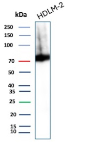
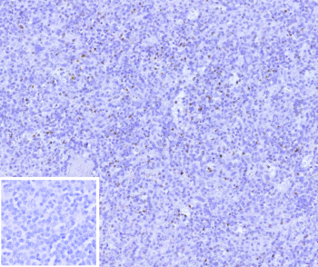
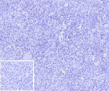
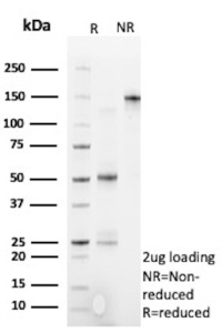
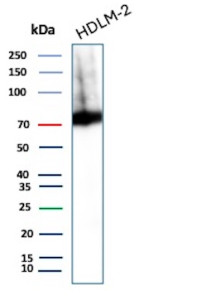
0.2 mg/ml in 1X PBS with 0.05% BSA, 0.05% sodium azide. Cytotoxic Lymphocyte Perforin Antibody recognizes Perforin, a calcium-dependent pore-forming protein encoded by the PRF1 gene. Perforin is a critical effector molecule of cytotoxic T lymphocytes and natural killer cells, where it is synthesized, stored in secretory granules, and released upon immune synapse formation. Following antigen recognition, Perforin is exocytosed and inserts into the membrane of target cells, forming transmembrane pores that allow granzymes to enter and initiate apoptosis. This tightly regulated mechanism is central to antiviral defense, tumor immunosurveillance, and elimination of intracellular pathogens.Perforin belongs to the membrane attack complex/perforin (MACPF) protein family and contains a conserved MACPF domain responsible for pore formation. The protein also includes calcium-binding regions that regulate its conformational change and membrane insertion. Under resting conditions, Perforin remains sequestered within cytotoxic granules to prevent unintended tissue damage. Upon activation of cytotoxic lymphocytes, controlled secretion ensures targeted killing of infected or transformed cells while minimizing collateral injury to surrounding tissues.The PRF1 gene is located on chromosome 10 and is predominantly expressed in activated CD8-positive T cells and natural killer cells. In lymphoid organs such as spleen, tonsil, and lymph node, Perforin-positive cells are typically scattered within germinal centers and interfollicular regions, reflecting the distribution of activated cytotoxic lymphocytes. Cytoplasmic granular staining is the expected localization pattern in immunohistochemistry due to storage within lytic granules.Genetic mutations in PRF1 are associated with familial hemophagocytic lymphohistiocytosis and other immune dysregulation syndromes, highlighting the essential role of Perforin in maintaining immune homeostasis. Altered expression levels have also been studied in cancer immunology, transplant rejection, and autoimmune disease research. Quantifying Perforin expression can provide insight into cytotoxic immune activity within tumor microenvironments or inflammatory lesions.This Cytotoxic Lymphocyte Perforin Antibody (clone PRF1/9681R) is a recombinant monoclonal antibody suitable for detecting PRF1 expression in research applications. A Cytotoxic Lymphocyte Perforin Antibody supports studies of cell-mediated cytotoxicity, immune activation status, and functional characterization of cytotoxic lymphocyte populations in normal and diseased tissues.
| Keywords: | Anti-PRF1, Anti-Cytolysin, Anti-Perforin-1, Anti-Lymphocyte pore-forming protein, Perforin Antibody / PRF1 |
| Supplier: | NSJ Bioreagents |
| Supplier-Nr: | V5975 |
Properties
| Application: | IHC, WB |
| Antibody Type: | Monoclonal |
| Clone: | PRF1/9681R |
| Conjugate: | No |
| Host: | Rabbit |
| Species reactivity: | human |
| Immunogen: | Recombinant fragment (around amino acids 400-555) of human Perforin-1 protein (exact sequence is proprietary) |
| Format: | Purified |
Database Information
| KEGG ID : | K07818 | Matching products |
| UniProt ID : | P14222 | Matching products |
| Gene ID : | GeneID 5551 | Matching products |
Handling & Safety
| Storage: | -20°C |
| Shipping: | -20°C (International: -20°C) |
Caution
Our products are for laboratory research use only: Not for administration to humans!
Our products are for laboratory research use only: Not for administration to humans!
Information about the product reference will follow.
more
You will get a certificate here
Viewed