Cookie preferences
This website uses cookies, which are necessary for the technical operation of the website and are always set. Other cookies, which increase the comfort when using this website, are used for direct advertising or to facilitate interaction with other websites and social networks, are only set with your consent.
Configuration
Technically required
These cookies are necessary for the basic functions of the shop.
"Allow all cookies" cookie
"Decline all cookies" cookie
CSRF token
Cookie preferences
Currency change
Customer-specific caching
FACT-Finder tracking
Individual prices
Selected shop
Session
Comfort functions
These cookies are used to make the shopping experience even more appealing, for example for the recognition of the visitor.
Note
Show the facebook fanpage in the right blod sidebar
Statistics & Tracking
Affiliate program
Conversion and usertracking via Google Tag Manager
Track device being used


| Item number | Size | Datasheet | Manual | SDS | Delivery time | Quantity | Price |
|---|---|---|---|---|---|---|---|
| NSJ-V6147-20UG | 20 µg | - | - |
3 - 10 business days* |
684.00€
|
||
| NSJ-V6147-100UG | 100 µg | - | - |
3 - 10 business days* |
1,160.00€
|
If you have any questions, please use our Contact Form.
You can also order by e-mail: info@biomol.com
Larger quantity required? Request bulk
You can also order by e-mail: info@biomol.com
Larger quantity required? Request bulk
Antibody in 1X PBS with 0.05% BSA, 0.05% sodium azide. Napsin A (NAPSA) is a lysosomal aspartic... more
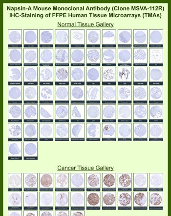
Product information "Anti-NAPSA / Napsin A, clone MSVA-112R"
Antibody in 1X PBS with 0.05% BSA, 0.05% sodium azide. Napsin A (NAPSA) is a lysosomal aspartic protease primarily expressed in lung alveolar epithelial cells and renal tubular epithelium, where it functions in protein processing within secretory and lysosomal compartments. NAPSA Antibody for IHC is widely used to detect Napsin A expression in formalin-fixed, paraffin-embedded tissues, supporting evaluation of epithelial differentiation and tumor classification. NAPSA antibody, also known as Napsin A antibody, is a well-established marker for lung adenocarcinoma and is frequently used in histopathology to distinguish primary lung tumors from metastatic carcinomas.Napsin A is predominantly localized to the cytoplasm in granular structures corresponding to lysosomes and secretory vesicles, reflecting its role in surfactant protein processing in type II pneumocytes. In normal tissues, expression is strongest in lung and kidney, with limited distribution in other epithelial cell types. In tumor settings, NAPSA expression is highly characteristic of lung adenocarcinoma, where strong cytoplasmic staining is observed in tumor epithelial cells, while most squamous cell carcinomas and non-pulmonary tumors lack expression, providing valuable diagnostic contrast.This NAPSA Antibody for IHC is uniquely positioned for detecting cytoplasmic Napsin A staining patterns in FFPE tissue sections, enabling clear visualization of epithelial tumor cell populations and supporting histological classification. The rabbit monoclonal recombinant format of clone MSVA-112R provides consistent performance and strong signal intensity, facilitating reliable detection of Napsin A in tissue microenvironments where precise cellular localization is critical.In immunohistochemistry applications, NAPSA antibody staining typically appears as granular cytoplasmic signal in lung adenocarcinoma cells, often with diffuse distribution throughout tumor regions. This staining pattern aligns with the intracellular localization of Napsin A within lysosomal and secretory compartments and supports its role as a marker of pulmonary epithelial differentiation. In contrast, stromal components and non-expressing tissues generally remain negative, enhancing contrast and interpretability in complex tissue sections.Beyond oncology, Napsin A expression also provides insight into epithelial cell biology and protease function within secretory pathways. Its involvement in surfactant protein maturation underscores its importance in lung physiology, while its restricted expression pattern makes it a useful marker for identifying specific epithelial cell populations. The NAPSA antibody therefore supports a wide range of research applications focused on tissue-specific protein expression, epithelial lineage tracing, and tumor microenvironment analysis.Overall, NAPSA antibody reagents are valuable tools for immunohistochemical detection of Napsin A, offering clear cytoplasmic staining patterns that aid in the identification of lung adenocarcinoma and the study of epithelial differentiation in both normal and diseased tissues.This antibody is part of a comprehensive NAPSA antibody collection developed to support Napsin A detection across IHC, WB, IF, and FACS applications in lung cancer and epithelial biology research.This antibody is also part of a broader collection of IHC antibodies validated by tissue microarray analysis, supporting consistent staining across normal and cancer tissues.
| Keywords: | Anti-NAPSA, Anti-Napsin-A, Anti-Napsin-1, Anti-TA01/TA02, Anti-Aspartyl protease 4, NAPSA Antibody / Napsin A |
| Supplier: | NSJ Bioreagents |
| Supplier-Nr: | V6147 |
Properties
| Application: | IHC |
| Antibody Type: | Monoclonal |
| Clone: | MSVA-112R |
| Conjugate: | No |
| Host: | Rabbit |
| Species reactivity: | human |
| Immunogen: | Recombinant human Napsin-A protein fragment (amino acids 189-299) (exact sequence is proprietary) |
| Format: | Purified |
Database Information
| KEGG ID : | K08565 | Matching products |
| UniProt ID : | O96009 | Matching products |
| Gene ID : | GeneID 9476 | Matching products |
Handling & Safety
| Storage: | -20°C |
| Shipping: | -20°C (International: -20°C) |
Caution
Our products are for laboratory research use only: Not for administration to humans!
Our products are for laboratory research use only: Not for administration to humans!
Information about the product reference will follow.
more
You will get a certificate here
Viewed