
Cookie preferences
This website uses cookies, which are necessary for the technical operation of the website and are always set. Other cookies, which increase the comfort when using this website, are used for direct advertising or to facilitate interaction with other websites and social networks, are only set with your consent.
Configuration
Technically required
These cookies are necessary for the basic functions of the shop.
"Allow all cookies" cookie
"Decline all cookies" cookie
CSRF token
Cookie preferences
Currency change
Customer-specific caching
FACT-Finder tracking
Individual prices
Selected shop
Session
Comfort functions
These cookies are used to make the shopping experience even more appealing, for example for the recognition of the visitor.
Note
Show the facebook fanpage in the right blod sidebar
Statistics & Tracking
Affiliate program
Conversion and usertracking via Google Tag Manager
Track device being used


| Item number | Size | Datasheet | Manual | SDS | Delivery time | Quantity | Price |
|---|---|---|---|---|---|---|---|
| NSJ-V5950-20UG | 20 µg | - | - |
7 - 12 business days* |
545.00€
|
||
| NSJ-V5950-100UG | 100 µg | - | - |
7 - 12 business days* |
1,080.00€
|
If you have any questions, please use our Contact Form.
You can also order by e-mail: info@biomol.com
Larger quantity required? Request bulk
You can also order by e-mail: info@biomol.com
Larger quantity required? Request bulk
Antibody in 1X PBS with 0.05% BSA, 0.05% sodium azide. Mucin 1 (MUC1), also known as Epithelial... more
Product information "Anti-MUC1 for IHC / Mucin 1, clone MSVA-672R"
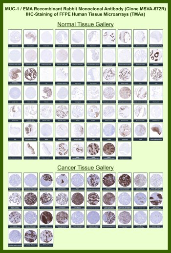
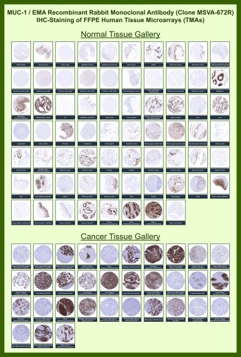
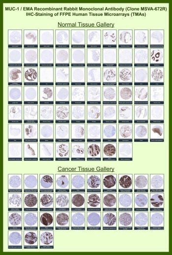
Antibody in 1X PBS with 0.05% BSA, 0.05% sodium azide. Mucin 1 (MUC1), also known as Epithelial Membrane Antigen (EMA), is a transmembrane glycoprotein expressed on the apical surface of epithelial cells, where it plays key roles in barrier formation, cell signaling, and epithelial polarity. MUC1 Antibody for IHC is optimized for detection of epithelial and tumor-associated expression in formalin-fixed, paraffin-embedded tissues, enabling high-contrast visualization of epithelial structures and carcinoma-associated staining patterns in histological sections.MUC1 antibody, also referred to as EMA antibody or MUC-1 antibody, is widely used in immunohistochemistry as a robust epithelial lineage marker with characteristic membranous and cytoplasmic staining. In normal tissues, MUC1 expression is predominantly localized to glandular and ductal epithelia, including breast, lung, gastrointestinal mucosa, pancreas, prostate, endometrium, and urothelium. In IHC staining, this polarized distribution frequently highlights luminal surfaces and apical membranes, providing clear delineation of epithelial architecture, while non-epithelial tissues such as muscle, connective tissue, and lymphoid compartments show minimal staining.Clone MSVA-672R is a recombinant rabbit monoclonal antibody developed for high-affinity and reproducible detection of MUC1 in FFPE samples. This clone produces strong membranous and cytoplasmic staining with low non-specific background under standard antigen retrieval conditions, enabling clear identification of epithelial cells and tumor structures. In Tissue Microarray (TMA) analysis, MUC1 Antibody for IHC demonstrates highly consistent staining across large panels of normal and cancer tissues, allowing side-by-side comparison of epithelial marker expression across hundreds of tissue cores within a single experiment.In normal tissue microarrays, MUC1 staining is prominently observed in glandular and ductal epithelial compartments, where strong HRP-DAB brown signal outlines luminal surfaces and epithelial cell borders. This pattern provides clear visualization of tissue organization and epithelial differentiation across multiple organ systems. Non-epithelial tissues, including stromal and mesenchymal compartments, are largely negative, reinforcing the specificity of MUC1 as an epithelial marker in immunohistochemistry.In cancer tissue microarrays, MUC1 Antibody for IHC reveals strong and often diffuse membranous and cytoplasmic staining in a wide range of epithelial malignancies, including breast carcinoma, lung adenocarcinoma, colorectal adenocarcinoma, pancreatic carcinoma, ovarian carcinoma, and urothelial carcinoma. Tumor cells frequently exhibit increased staining intensity and loss of apical polarization compared to normal tissues, reflecting altered MUC1 distribution during malignant transformation. This staining pattern is particularly valuable for identifying epithelial-derived tumor cells, assessing tumor differentiation, and distinguishing carcinoma cells from surrounding stromal components in complex tissue environments.The robust and reproducible performance of clone MSVA-672R in TMA-based immunohistochemistry supports its application in cancer research, tumor profiling, and biomarker validation studies. MUC1 Antibody for IHC enables reliable detection of epithelial markers in FFPE tissues and is well suited for high-throughput tissue microarray analysis, comparative pathology, and evaluation of epithelial tumor biology across diverse tissue types.This antibody is also part of a broader collection of IHC antibodies validated by tissue microarray analysis, supporting consistent staining across normal and cancer tissues.
| Keywords: | Anti-MUC1, Anti-PEMT, Anti-H23AG, Anti-Mucin-1, Anti-Episialin, Anti-Cancer antigen 15-3, Anti-Krebs von den Lungen-6, Anti-Tumor-associated mucin, Anti-Carcinoma-associated mucin, Anti-Polymorphic epithelial mucin, Anti-Peanut-reactive urinary mucin, Ant |
| Supplier: | NSJ Bioreagents |
| Supplier-Nr: | V5950 |
Properties
| Application: | IHC |
| Antibody Type: | Monoclonal |
| Clone: | MSVA-672R |
| Conjugate: | No |
| Host: | Rabbit |
| Species reactivity: | human |
| Immunogen: | Recombinant human Mucin 1 protein |
| Format: | Purified |
Database Information
| KEGG ID : | K06568 | Matching products |
| UniProt ID : | P15941 | Matching products |
| Gene ID : | GeneID 4582 | Matching products |
Handling & Safety
| Storage: | -20°C |
| Shipping: | -20°C (International: -20°C) |
Caution
Our products are for laboratory research use only: Not for administration to humans!
Our products are for laboratory research use only: Not for administration to humans!
Information about the product reference will follow.
more
You will get a certificate here
Viewed