Cookie preferences
This website uses cookies, which are necessary for the technical operation of the website and are always set. Other cookies, which increase the comfort when using this website, are used for direct advertising or to facilitate interaction with other websites and social networks, are only set with your consent.
Configuration
Technically required
These cookies are necessary for the basic functions of the shop.
"Allow all cookies" cookie
"Decline all cookies" cookie
CSRF token
Cookie preferences
Currency change
Customer-specific caching
FACT-Finder tracking
Individual prices
Selected shop
Session
Comfort functions
These cookies are used to make the shopping experience even more appealing, for example for the recognition of the visitor.
Note
Show the facebook fanpage in the right blod sidebar
Statistics & Tracking
Affiliate program
Conversion and usertracking via Google Tag Manager
Track device being used


| Item number | Size | Datasheet | Manual | SDS | Delivery time | Quantity | Price |
|---|---|---|---|---|---|---|---|
| NSJ-V6098-20UG | 20 µg | - | - |
3 - 10 business days* |
684.00€
|
||
| NSJ-V6098-100UG | 100 µg | - | - |
3 - 10 business days* |
1,160.00€
|
If you have any questions, please use our Contact Form.
You can also order by e-mail: info@biomol.com
Larger quantity required? Request bulk
You can also order by e-mail: info@biomol.com
Larger quantity required? Request bulk
Antibody in 1X PBS with 0.05% BSA, 0.05% sodium azide. Myeloperoxidase (MPO) is a heme-containing... more
Product information "Anti-MPO / Myeloperoxidase, clone MSVA-692M"
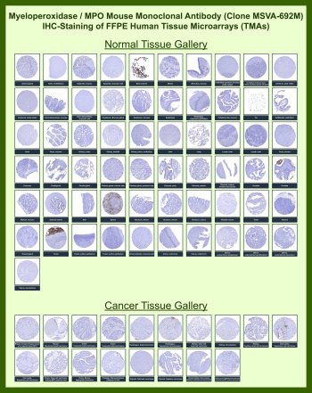
Antibody in 1X PBS with 0.05% BSA, 0.05% sodium azide. Myeloperoxidase (MPO) is a heme-containing peroxidase enzyme encoded by the MPO gene and predominantly expressed in neutrophils and other cells of the myeloid lineage. Myeloperoxidase Antibody for IHC / MPO Immunohistochemistry Antibody (clone MSVA-692M) enables visualization of MPO expression in formalin-fixed, paraffin-embedded tissue sections using immunohistochemistry. This approach allows researchers to identify MPO-positive myeloid cells within the histological architecture of normal tissues and tumors.MPO antibody, also referred to as Myeloperoxidase antibody or myeloid peroxidase antibody in the literature, detects an abundant cytoplasmic enzyme localized within the azurophilic granules of neutrophils and other granulocytic cells. Because of this restricted expression pattern, immunohistochemistry staining with Myeloperoxidase Antibody for IHC highlights granulocytes and related myeloid lineage cells in tissue sections. The staining pattern typically appears as granular cytoplasmic labeling corresponding to MPO-containing granules.Immunohistochemistry analysis with MPO antibodies is widely used in hematopathology and cancer research to identify cells of myeloid origin. MPO immunohistochemistry staining can distinguish neutrophil lineage cells and is frequently used to evaluate inflammatory infiltrates or characterize myeloid differentiation in tumor or hematologic specimens. Cytoplasmic MPO staining within granulocytes provides a clear histological marker of myeloid cell populations.Immunohistochemistry also allows investigators to examine MPO-positive cells within the context of tissue architecture. In normal tissues, MPO-positive granulocytes may be observed within bone marrow or circulating within blood-rich organs. In tumor samples or inflammatory tissues, immunohistochemistry staining may highlight infiltrating neutrophils or other MPO-expressing cells present within the tumor microenvironment.Myeloperoxidase Antibody for IHC (clone MSVA-692M) has been evaluated by immunohistochemistry across a wide range of normal and cancer tissues using tissue microarray analysis. The immunohistochemistry staining patterns observed with this antibody align with Myeloperoxidase / MPO expression data reported in the Human Protein Atlas, supporting its use for histological detection of MPO-expressing cells. This antibody therefore supports immunohistochemistry-based visualization of MPO-positive myeloid cells in tissue sections.
| Keywords: | Anti-MPO, Anti-Myeloperoxidase, MPO Antibody / Myeloperoxidase |
| Supplier: | NSJ Bioreagents |
| Supplier-Nr: | V6098 |
Properties
| Application: | IHC |
| Antibody Type: | Monoclonal |
| Clone: | MSVA-692M |
| Conjugate: | No |
| Host: | Mouse |
| Species reactivity: | human |
| Immunogen: | Recombinant fragment of human MPO protein (exact sequence is proprietary) |
| Format: | Purified |
Database Information
| KEGG ID : | K10789 | Matching products |
| UniProt ID : | P05164 | Matching products |
| Gene ID : | GeneID 4353 | Matching products |
Handling & Safety
| Storage: | -20°C |
| Shipping: | -20°C (International: -20°C) |
Caution
Our products are for laboratory research use only: Not for administration to humans!
Our products are for laboratory research use only: Not for administration to humans!
Information about the product reference will follow.
more
You will get a certificate here
Viewed