
Cookie preferences
This website uses cookies, which are necessary for the technical operation of the website and are always set. Other cookies, which increase the comfort when using this website, are used for direct advertising or to facilitate interaction with other websites and social networks, are only set with your consent.
Configuration
Technically required
These cookies are necessary for the basic functions of the shop.
"Allow all cookies" cookie
"Decline all cookies" cookie
CSRF token
Cookie preferences
Currency change
Customer-specific caching
FACT-Finder tracking
Individual prices
Selected shop
Session
Comfort functions
These cookies are used to make the shopping experience even more appealing, for example for the recognition of the visitor.
Note
Show the facebook fanpage in the right blod sidebar
Statistics & Tracking
Affiliate program
Conversion and usertracking via Google Tag Manager
Track device being used


| Item number | Size | Datasheet | Manual | SDS | Delivery time | Quantity | Price |
|---|---|---|---|---|---|---|---|
| NSJ-FY12787 | 100 µg | - | - |
3 - 10 business days* |
790.00€
|
If you have any questions, please use our Contact Form.
You can also order by e-mail: info@biomol.com
Larger quantity required? Request bulk
You can also order by e-mail: info@biomol.com
Larger quantity required? Request bulk
Adding 0.2 ml of distilled water will yield a concentration of 500 ug/ml. MICAL3 antibody detects... more
Product information "Anti-MICAL3 / Molecule interacting with CasL protein 3"
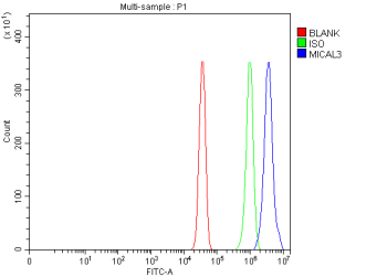
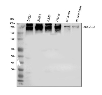
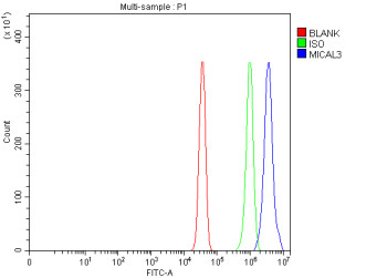
Adding 0.2 ml of distilled water will yield a concentration of 500 ug/ml. MICAL3 antibody detects Microtubule-associated monooxygenase, calponin, and LIM domain-containing protein 3, an actin-modifying enzyme that regulates cytoskeletal organization and vesicle trafficking. Encoded by the MICAL3 gene on chromosome 22q11.21, this multidomain oxidoreductase functions downstream of Rab GTPases to control exocytosis and vesicle docking at the plasma membrane. MICAL3 is part of the MICAL protein family, characterized by an N-terminal flavoprotein monooxygenase domain, calponin homology domain, LIM domain, and coiled-coil regions that mediate interactions with cytoskeletal and vesicular components.MICAL3 catalyzes F-actin oxidation, leading to filament disassembly and cytoskeletal remodeling required for vesicle fusion. It interacts with Rab8A and Rab13, forming complexes that regulate polarized exocytosis and membrane trafficking in epithelial cells. MICAL3 is localized to vesicular membranes and actin-rich regions of the cytoplasm, where it coordinates actin depolymerization and vesicle release. Its enzymatic activity is dependent on flavin adenine dinucleotide (FAD) and modulates actin dynamics through redox reactions on specific methionine residues.The MICAL3 antibody is used in cell biology, neurobiology, and membrane trafficking research to study cytoskeletal regulation and exocytosis. Western blot analysis identifies a 230 kilodalton band corresponding to MICAL3, while immunofluorescence reveals punctate cytoplasmic staining in vesicle-associated regions. This antibody enables investigation of actin redox control, vesicle tethering, and neuronal growth cone dynamics.MICAL3 has been implicated in axonal transport, endocytosis, and cancer cell migration. Its deregulation may contribute to disorders involving vesicle trafficking or cytoskeletal disorganization. The MICAL3 antibody provides a validated tool for studying redox-regulated actin remodeling and membrane dynamics. NSJ Bioreagents offers this antibody optimized for western blotting and flow cytometry, ensuring high sensitivity and reproducibility in studies of cytoskeletal and vesicular processes.
| Keywords: | Anti-MICAL3, Anti-[F-actin]-monooxygenase MICAL3, Anti-Molecule interacting with CasL protein 3, MICAL3 Antibody / Molecule interacting with CasL protein 3 |
| Supplier: | NSJ Bioreagents |
| Supplier-Nr: | FY12787 |
Properties
| Application: | WB, FC, ELISA |
| Antibody Type: | Polyclonal |
| Conjugate: | No |
| Host: | Rabbit |
| Species reactivity: | human, mouse, rat |
| Immunogen: | E.coli-derived human MICAL3 recombinant protein (Position: K458-L1592) |
| Format: | Purified |
Database Information
| KEGG ID : | K19947 | Matching products |
| UniProt ID : | Q7RTP6 | Matching products |
| Gene ID : | GeneID 57553 | Matching products |
Handling & Safety
| Storage: | +4°C |
| Shipping: | +4°C (International: +4°C) |
Caution
Our products are for laboratory research use only: Not for administration to humans!
Our products are for laboratory research use only: Not for administration to humans!
Information about the product reference will follow.
more
You will get a certificate here
Viewed