Cookie preferences
This website uses cookies, which are necessary for the technical operation of the website and are always set. Other cookies, which increase the comfort when using this website, are used for direct advertising or to facilitate interaction with other websites and social networks, are only set with your consent.
Configuration
Technically required
These cookies are necessary for the basic functions of the shop.
"Allow all cookies" cookie
"Decline all cookies" cookie
CSRF token
Cookie preferences
Currency change
Customer-specific caching
FACT-Finder tracking
Individual prices
Selected shop
Session
Comfort functions
These cookies are used to make the shopping experience even more appealing, for example for the recognition of the visitor.
Note
Show the facebook fanpage in the right blod sidebar
Statistics & Tracking
Affiliate program
Conversion and usertracking via Google Tag Manager
Track device being used


| Item number | Size | Datasheet | Manual | SDS | Delivery time | Quantity | Price |
|---|---|---|---|---|---|---|---|
| NSJ-FY12964 | 100 µl | - | - |
3 - 10 business days* |
790.00€
|
If you have any questions, please use our Contact Form.
You can also order by e-mail: info@biomol.com
Larger quantity required? Request bulk
You can also order by e-mail: info@biomol.com
Larger quantity required? Request bulk
Rabbit IgG in phosphate buffered saline, pH 7.4, 150mM NaCl, 0.02% sodium azide and 50% glycerol,... more
Product information "Anti-Ki-67 / MKI67, clone 31K02"
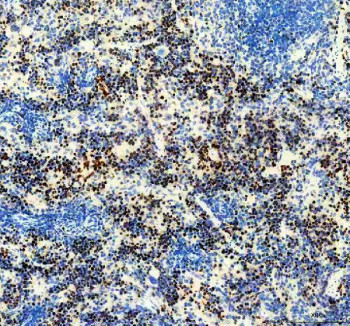
Rabbit IgG in phosphate buffered saline, pH 7.4, 150mM NaCl, 0.02% sodium azide and 50% glycerol, 0.4-0.5mg/ml BSA. Ki-67 antibody detects the protein Ki-67, encoded by the MKI67 gene. Ki-67 is a nuclear protein expressed in proliferating cells and is one of the most widely used markers for cellular proliferation in research and clinical diagnostics. This protein is absent in resting cells (G0) but present during all active phases of the cell cycle, including G1, S, G2, and mitosis. Ki-67 antibody provides researchers with an essential tool to measure cell proliferation rates, assess tumor aggressiveness, and evaluate treatment responses in cancer studies. Its widespread use in histopathology reflects its reliability as a biomarker for growth activity.Ki-67 is involved in ribosomal RNA transcription and organization of the nucleolus during interphase. During mitosis, it localizes to the perichromosomal layer, forming a structural scaffold that helps protect chromosomes and maintain proper segregation. Studies using Ki-67 antibody have revealed that depletion of this protein disrupts chromosomal condensation and segregation, underscoring its functional roles beyond serving as a proliferation marker. These insights have expanded its importance from being a passive marker of division to an active participant in cell cycle regulation.In cancer biology, Ki-67 is one of the most important clinical biomarkers. Tumor samples are routinely stained with Ki-67 antibody to determine the proliferation index, which correlates with tumor grade, aggressiveness, and prognosis. High Ki-67 labeling indices are observed in fast-growing tumors such as breast, prostate, and brain cancers, and are associated with poor outcomes. Conversely, lower Ki-67 indices are linked to slower-growing neoplasms and better survival rates. Pathologists use Ki-67 antibody immunohistochemistry to quantify nuclear staining percentages in tumor biopsies, providing valuable prognostic information.Beyond oncology, Ki-67 antibody is applied in regenerative biology and developmental studies. Since this protein marks actively dividing cells, it is used to track stem cell proliferation, tissue regeneration, and embryonic development. Immunofluorescence assays with Ki-67 antibody help identify proliferative niches in tissues such as bone marrow, gut, and brain. Flow cytometry can also measure Ki-67 expression at the single-cell level, making it useful in studies of immune activation, hematopoietic stem cell dynamics, and vaccine responses.By supplying validated Ki-67 antibody reagents, NSJ Bioreagents enables researchers to study both the biological roles of this protein and its diagnostic utility. The ability to detect proliferating cells across experimental systems makes Ki-67 antibody indispensable in cancer research, clinical pathology, and developmental biology.
| Keywords: | Anti-MKI67, Anti-Proliferation marker protein Ki-67, Anti-Antigen identified by monoclonal antibody Ki-67, Ki-67 Antibody / MKI67 |
| Supplier: | NSJ Bioreagents |
| Supplier-Nr: | FY12964 |
Properties
| Application: | IHC, IF, ICC/IF |
| Antibody Type: | Monoclonal |
| Clone: | 31K02 |
| Conjugate: | No |
| Host: | Rabbit |
| Species reactivity: | human, mouse, rat |
| Immunogen: | Recombinant protein with human Ki67 (Position: K2860-I3256) |
| Format: | Affinity Purified |
Database Information
| KEGG ID : | K17582 | Matching products |
| UniProt ID : | P46013 | Matching products |
| Gene ID : | GeneID 4288 | Matching products |
Handling & Safety
| Storage: | -20°C |
| Shipping: | -20°C (International: -20°C) |
Caution
Our products are for laboratory research use only: Not for administration to humans!
Our products are for laboratory research use only: Not for administration to humans!
Information about the product reference will follow.
more
You will get a certificate here
Viewed