
Cookie preferences
This website uses cookies, which are necessary for the technical operation of the website and are always set. Other cookies, which increase the comfort when using this website, are used for direct advertising or to facilitate interaction with other websites and social networks, are only set with your consent.
Configuration
Technically required
These cookies are necessary for the basic functions of the shop.
"Allow all cookies" cookie
"Decline all cookies" cookie
CSRF token
Cookie preferences
Currency change
Customer-specific caching
FACT-Finder tracking
Individual prices
Selected shop
Session
Comfort functions
These cookies are used to make the shopping experience even more appealing, for example for the recognition of the visitor.
Note
Show the facebook fanpage in the right blod sidebar
Statistics & Tracking
Affiliate program
Conversion and usertracking via Google Tag Manager
Track device being used



| Item number | Size | Datasheet | Manual | SDS | Delivery time | Quantity | Price |
|---|---|---|---|---|---|---|---|
| NSJ-V6097-20UG | 20 µg | - | - |
3 - 10 business days* |
684.00€
|
||
| NSJ-V6097-100UG | 100 µg | - | - |
3 - 10 business days* |
1,265.00€
|
If you have any questions, please use our Contact Form.
You can also order by e-mail: info@biomol.com
Larger quantity required? Request bulk
You can also order by e-mail: info@biomol.com
Larger quantity required? Request bulk
Antibody in 1X PBS with 0.05% BSA, 0.05% sodium azide. Ki-67 antigen (MKI67) is a nuclear... more
Product information "Anti-Ki-67 / Marker of proliferation Ki-67, clone MSVA-267M"
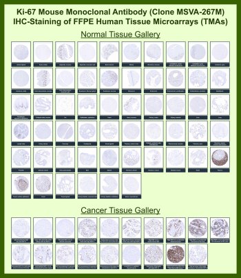
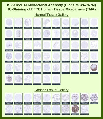
Antibody in 1X PBS with 0.05% BSA, 0.05% sodium azide. Ki-67 antigen (MKI67) is a nuclear proliferation-associated protein encoded by the MKI67 gene and is widely used as a marker of actively cycling cells in normal and neoplastic tissues. Ki-67 Antibody for IHC / Ki-67 IHC Antibody is developed for immunohistochemistry detection of this well-established proliferation marker in formalin-fixed, paraffin-embedded tissue sections. Because Ki-67 expression is closely linked to cell cycle activity and is largely absent in quiescent cells, immunohistochemistry staining provides a practical way to visualize proliferative compartments directly within preserved tissue architecture.Ki-67 antibody, also referred to as Ki67 antibody, MKI67 antibody, or Antigen KI-67 antibody in the literature, detects a protein that localizes predominantly to the nucleus during active phases of the cell cycle. In immunohistochemistry studies, Ki-67 staining typically appears as brown nuclear chromogenic signal in proliferating cells, allowing clear distinction between cycling and non-cycling populations. This makes Ki-67 Antibody for IHC especially useful for assessing germinal centers, basal proliferative zones, intestinal crypt compartments, and tumor cell populations with increased growth fraction.The value of immunohistochemistry for Ki-67 lies in its ability to combine proliferation assessment with tissue morphology. In normal tissues, nuclear Ki-67 staining is usually confined to expected proliferative compartments, while differentiated or resting cells show little to no signal. In tumors, Ki-67 IHC antibody staining often highlights variable fractions of proliferating malignant nuclei, helping researchers examine growth patterns across carcinomas, lymphomas, sarcomas, and other neoplasms. The intensity and distribution of nuclear staining can therefore provide useful visual context when comparing different tumor areas or tissue types in research settings.Ki-67 is expressed during G1, S, G2, and mitotic phases, but is generally not detected in resting G0 cells. At the subcellular level, the protein associates with the nucleus and shows characteristic changes in distribution during mitosis. This biology explains why immunohistochemistry staining with Ki-67 antibodies is widely used to identify proliferative activity rather than lineage alone. In FFPE sections, the expected finding is selective nuclear labeling rather than cytoplasmic or membranous signal, which helps support straightforward microscopic interpretation.Ki-67 Antibody for IHC / Ki-67 IHC Antibody provides a valuable tool for immunohistochemistry-based evaluation of proliferating cells in both normal and cancer tissues. By highlighting nuclear expression of Ki-67 / MKI67 within intact histologic context, this antibody supports studies of tumor biology, tissue turnover, growth fraction, and cellular proliferation in pathology-oriented research.This antibody is also part of a broader collection of IHC antibodies validated by tissue microarray analysis, supporting consistent staining across normal and cancer tissues.
| Keywords: | Anti-MKI67, Anti-Proliferation marker protein Ki-67, Anti-Antigen identified by monoclonal antibody Ki-67, Ki-67 Antibody / Marker of proliferation Ki-67 |
| Supplier: | NSJ Bioreagents |
| Supplier-Nr: | V6097 |
Properties
| Application: | IHC |
| Antibody Type: | Monoclonal |
| Clone: | MSVA-267M |
| Conjugate: | No |
| Host: | Mouse |
| Species reactivity: | human |
| Immunogen: | Recombinant human Ki67 protein fragment (around amino acids 2293-2478) (exact sequence is proprietary) |
| Format: | Purified |
Database Information
| KEGG ID : | K17582 | Matching products |
| UniProt ID : | P46013 | Matching products |
| Gene ID : | GeneID 4288 | Matching products |
Handling & Safety
| Storage: | -20°C |
| Shipping: | -20°C (International: -20°C) |
Caution
Our products are for laboratory research use only: Not for administration to humans!
Our products are for laboratory research use only: Not for administration to humans!
Information about the product reference will follow.
more
You will get a certificate here
Viewed