
Cookie preferences
This website uses cookies, which are necessary for the technical operation of the website and are always set. Other cookies, which increase the comfort when using this website, are used for direct advertising or to facilitate interaction with other websites and social networks, are only set with your consent.
Configuration
Technically required
These cookies are necessary for the basic functions of the shop.
"Allow all cookies" cookie
"Decline all cookies" cookie
CSRF token
Cookie preferences
Currency change
Customer-specific caching
FACT-Finder tracking
Individual prices
Selected shop
Session
Comfort functions
These cookies are used to make the shopping experience even more appealing, for example for the recognition of the visitor.
Note
Show the facebook fanpage in the right blod sidebar
Statistics & Tracking
Affiliate program
Conversion and usertracking via Google Tag Manager
Track device being used


| Item number | Size | Datasheet | Manual | SDS | Delivery time | Quantity | Price |
|---|---|---|---|---|---|---|---|
| NSJ-V6144-20UG | 20 µg | - | - |
3 - 10 business days* |
684.00€
|
||
| NSJ-V6144-100UG | 100 µg | - | - |
3 - 10 business days* |
1,265.00€
|
If you have any questions, please use our Contact Form.
You can also order by e-mail: info@biomol.com
Larger quantity required? Request bulk
You can also order by e-mail: info@biomol.com
Larger quantity required? Request bulk
Antibody in 1X PBS with 0.05% BSA, 0.05% sodium azide. Cluster of Differentiation 163 (CD163),... more
Product information "Anti-CD163, clone MSVA-163M"
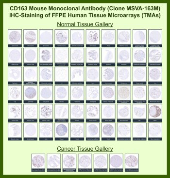
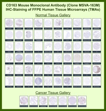
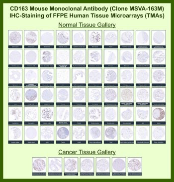
Antibody in 1X PBS with 0.05% BSA, 0.05% sodium azide. Cluster of Differentiation 163 (CD163), encoded by the CD163 gene, is a hemoglobin scavenger receptor expressed predominantly on monocytes and tissue-resident macrophages. CD163 is widely recognized as a marker of alternatively activated macrophages and plays a central role in clearance of hemoglobin-haptoglobin complexes, regulation of inflammation, and tissue remodeling. CD163 expression is particularly enriched in macrophage populations associated with anti-inflammatory and immunoregulatory functions and is commonly used to identify M2-like macrophages in tissue sections.CD163 Antibody for IHC, also referred to as CD163 immunohistochemistry antibody or macrophage marker antibody in the literature, is specifically optimized for detecting CD163-positive cells in formalin-fixed, paraffin-embedded tissues. This CD163 Antibody for IHC / CD163 Immunohistochemistry Antibody is uniquely positioned for high-confidence identification of macrophage populations within intact tissue architecture, enabling detailed evaluation of immune cell infiltration and spatial distribution. Clone MSVA-163M antibody has been tested on human tissue microarrays (TMA), supporting consistent staining performance across a broad range of normal and cancer tissues and reinforcing its suitability for comparative tissue analysis.In immunohistochemistry applications, CD163 is typically observed as strong membranous and cytoplasmic HRP-DAB brown staining in macrophages localized within stromal regions, perivascular niches, and sites of active inflammation. Tissue microarray (TMA) analysis demonstrates robust and reproducible labeling of macrophage populations across multiple organ systems, including spleen, liver, lung, and lymphoid tissues, while most non-hematopoietic parenchymal cells remain negative. This consistent staining pattern supports reliable identification of macrophage lineage cells across diverse tissue contexts.Within tumor tissues, CD163-positive tumor-associated macrophages are frequently enriched in the tumor microenvironment, where they localize around tumor nests, invasive fronts, and vascular structures. TMA-based evaluation highlights variability in macrophage density and distribution across different tumor types, providing insight into tumor microenvironment composition and immune infiltration patterns. These macrophages are associated with immunosuppressive signaling, extracellular matrix remodeling, and promotion of tumor growth and angiogenesis.A major advantage of using a CD163 antibody for IHC is its ability to distinguish macrophage subsets with anti-inflammatory and pro-tumorigenic characteristics. CD163-positive macrophages are often correlated with poor prognosis in multiple cancer types, including breast, lung, and colorectal carcinomas. Tissue microarray analysis further enables high-throughput comparison of macrophage infiltration across large sample sets, supporting biomarker studies and translational research.CD163 expression is regulated by cytokines such as IL10, glucocorticoids, and other anti-inflammatory mediators, and is upregulated in conditions associated with chronic inflammation, tissue repair, and immune modulation. Detection of CD163 by immunohistochemistry therefore provides insight into macrophage activation states and the balance between pro-inflammatory and anti-inflammatory signaling within tissues.In addition to its role in hemoglobin clearance, CD163 contributes to modulation of oxidative stress and cytokine signaling pathways that support resolution of inflammation and maintenance of tissue homeostasis. These functional roles make CD163 a valuable marker for studying macrophage biology in both physiological and pathological contexts.The CD163 gene is located on chromosome 12p13 and encodes a transmembrane protein belonging to the scavenger receptor cysteine-rich superfamily. Its restricted expression pattern and strong association with macrophage lineage cells make it a highly specific and reliable marker for immunohistochemical detection.This CD163 antibody for IHC is suitable for detecting CD163 expression in formalin-fixed tissue sections, with demonstrated performance in tissue microarray (TMA) analysis supporting studies of tumor microenvironment composition, macrophage infiltration, and inflammatory disease processes.This antibody is part of a broader collection of CD Antibodies designed for detecting immune cell markers and profiling leukocyte populations across tissue-based and cellular assays.This antibody is also part of a broader collection of IHC antibodies validated by tissue microarray analysis, supporting consistent staining across normal and cancer tissues.
| Keywords: | Anti-CD163, Anti-Hemoglobin scavenger receptor, Anti-Scavenger receptor cysteine-rich type 1 protein M130, CD163 Antibody |
| Supplier: | NSJ Bioreagents |
| Supplier-Nr: | V6144 |
Properties
| Application: | IHC |
| Antibody Type: | Monoclonal |
| Clone: | MSVA-163M |
| Conjugate: | No |
| Host: | Mouse |
| Species reactivity: | human |
| Immunogen: | Recombinant fragment of human CD163 protein (around amino acids 43-196) (exact sequence is proprietary) |
| Format: | Purified |
Database Information
| KEGG ID : | K06545 | Matching products |
| UniProt ID : | Q86VB7 | Matching products |
| Gene ID : | GeneID 9332 | Matching products |
Handling & Safety
| Storage: | -20°C |
| Shipping: | -20°C (International: -20°C) |
Caution
Our products are for laboratory research use only: Not for administration to humans!
Our products are for laboratory research use only: Not for administration to humans!
Information about the product reference will follow.
more
You will get a certificate here
Viewed