
Cookie preferences
This website uses cookies, which are necessary for the technical operation of the website and are always set. Other cookies, which increase the comfort when using this website, are used for direct advertising or to facilitate interaction with other websites and social networks, are only set with your consent.
Configuration
Technically required
These cookies are necessary for the basic functions of the shop.
"Allow all cookies" cookie
"Decline all cookies" cookie
CSRF token
Cookie preferences
Currency change
Customer-specific caching
FACT-Finder tracking
Individual prices
Selected shop
Session
Comfort functions
These cookies are used to make the shopping experience even more appealing, for example for the recognition of the visitor.
Note
Show the facebook fanpage in the right blod sidebar
Statistics & Tracking
Affiliate program
Conversion and usertracking via Google Tag Manager
Track device being used




Please complete the Material Transfer Agreement (MTA) and submit it along with your order. For all commercial applications, please complete the Intended Use Form.
| Item number | Size | Datasheet | Manual | SDS | Delivery time | Quantity | Price |
|---|---|---|---|---|---|---|---|
| CYT-300414 | 1 each | - |
3 - 8 business days* |
800.00€
|
If you have any questions, please use our Contact Form.
You can also order by e-mail: info@biomol.com
Larger quantity required? Request bulk
You can also order by e-mail: info@biomol.com
Larger quantity required? Request bulk
Categories: Kidney cancer cell lines Description: The Wilms3 cell line was established from a... more
Product information "Wilms3 Cells"
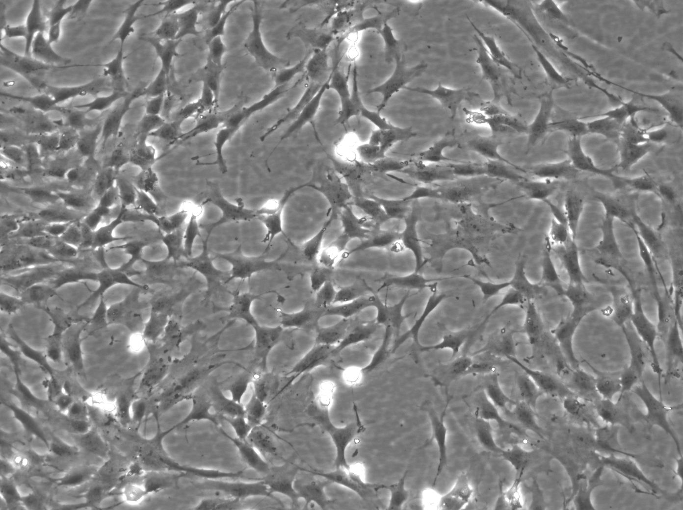
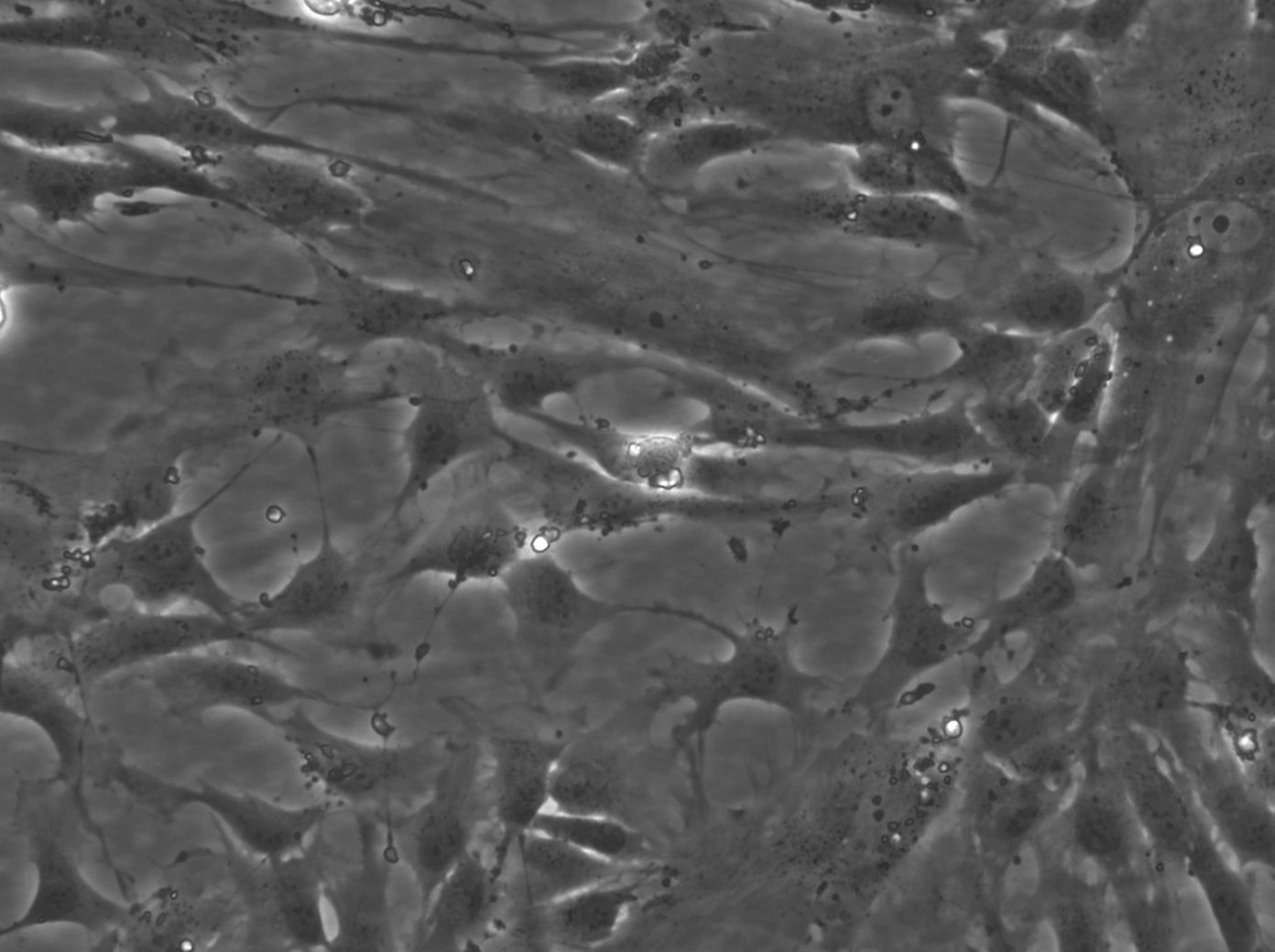
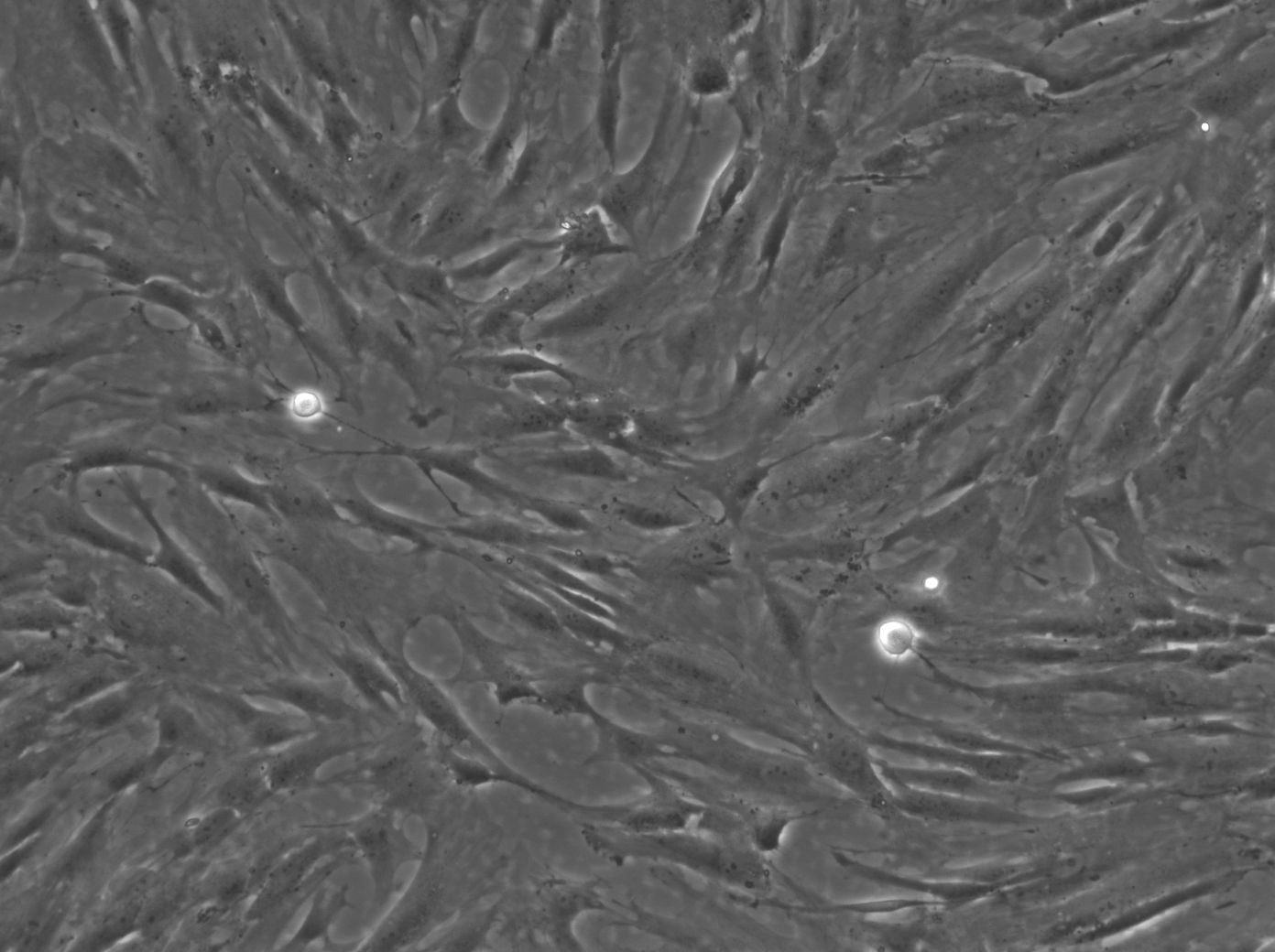
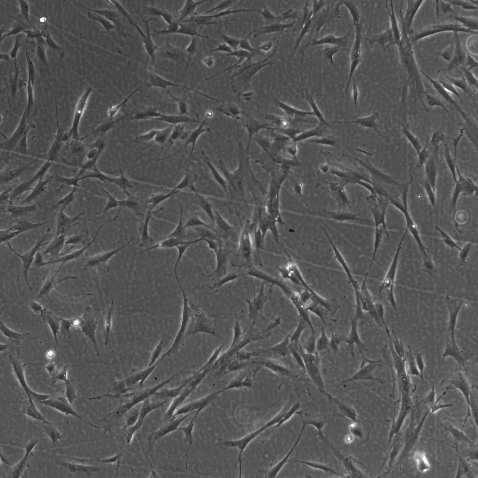
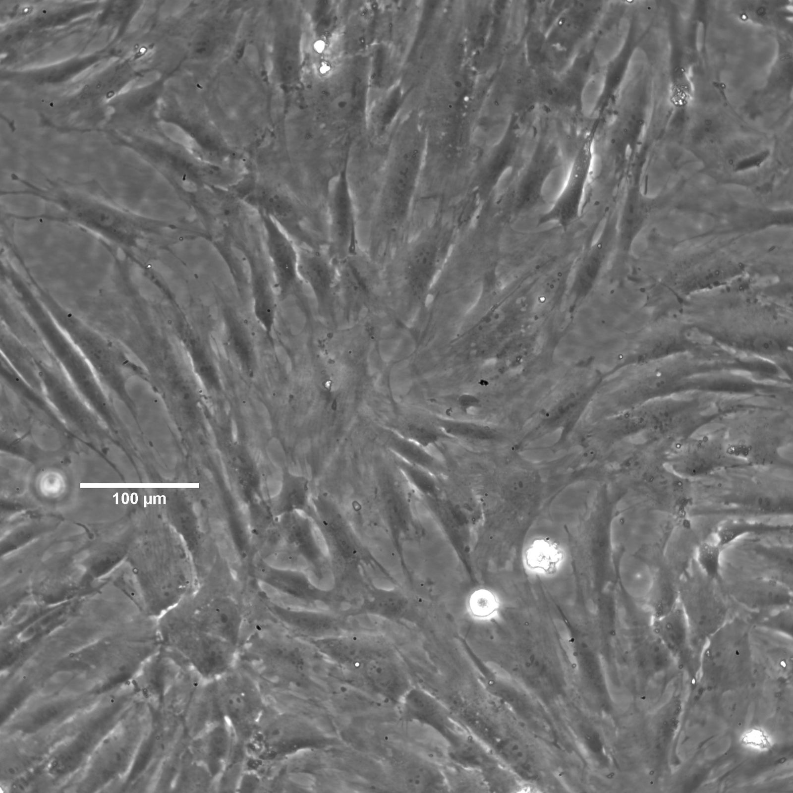
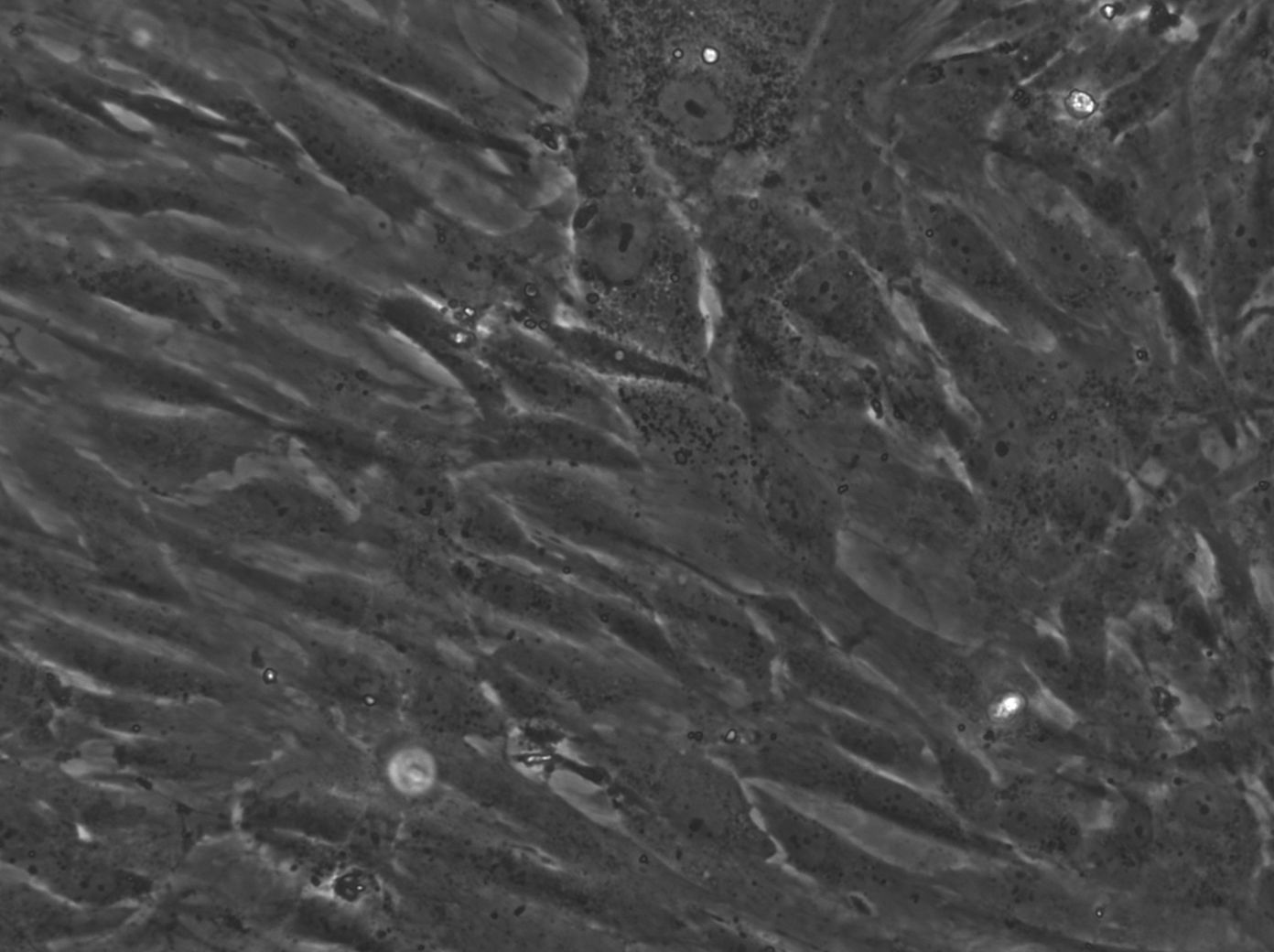
Categories: Kidney cancer cell lines Description: The Wilms3 cell line was established from a primary Wilms tumor in a pediatric patient, characterized by a somatic WT1 mutation. Unlike many other Wilms tumor cell lines, Wilms3 harbors a heterozygous frameshift mutation in the WT1 gene (c.1293-1294insA, p.V432SfsX87), leading to the production of a truncated WT1 protein. This partial loss of WT1 function is associated with the development of tumors that display a stromal or mesenchymal phenotype. However, the WT1 mutation in Wilms3 is not homozygous, which adds complexity to its study, as it retains some WT1 function that can influence tumor biology differently compared to cell lines with complete WT1 loss. Wilms3 also carries a mutation in the CTNNB1 gene, specifically affecting threonine 41 (p.T41A), which plays a critical role in the Wnt signaling pathway. This mutation stabilizes beta-Catenin, preventing its degradation and leading to the constitutive activation of the Wnt pathway. The persistent activation of Wnt signaling drives cell proliferation and contributes to tumorigenesis in Wilms3, making it a key model for studying the impact of CTNNB1 mutations in the context of a partially functional WT1 background. Phenotypically, Wilms3 cells exhibit a mesenchymal-like morphology, expressing vimentin and lacking cytokeratin, consistent with the stromal characteristics observed in the original tumor. These cells show limited differentiation potential, with the ability to undergo some mesenchymal differentiation under specific conditions. Proteomic analyses of Wilms3 have revealed the activation of several receptor tyrosine kinases (RTKs), including PDGFRbeta and AXL, which support cell survival and proliferation. Additionally, downstream signaling pathways such as MAPK and PI3K/AKT are activated, reinforcing the malignant properties of Wilms3 cells. One unique aspect of Wilms3 is its partial WT1 functionality, which provides a distinct perspective on how WT1 mutations contribute to Wilms tumor biology when the mutation is not complete. The interplay between WT1 and Wnt signaling in Wilms3 offers a valuable opportunity to study the nuanced roles these pathways play in tumor development. Overall, Wilms3 serves as an important model for investigating the molecular mechanisms underlying Wilms tumor in the presence of partial WT1 loss and constitutive Wnt pathway activation. Organism: Human Tissue: Kidney Disease: Wilms tumor Age: 11-12 months Gender: Male Ethnicity: Caucasian Morphology: Spindle-shaped Cell Type: Wilms cells Growth Properties: Adherent Citation: Wilms3 (Cytion catalog number 300414) Biosafety Level: 1 Ncbi_ Taxid: 9606.0 Cellosaurus Accession: CVCL_A5SF Mutational Profile: WT1 mutation status: homozygous c.1293-1294insA, p.V432fsx87, LOH: 11p11-11pter, CTNNB1 mutation status: wild type Culture Medium: MSCGM kit (from Lonza) Dissociation Reagent: Accutase Subculturing: Remove the old medium from the adherent cells and wash them with PBS that lacks calcium and magnesium. For T25 flasks, use 3-5 ml of PBS, and for T75 flasks, use 5-10 ml. Then, cover the cells completely with Accutase, using 1-2 ml for T25 flasks and 2.5 ml for T75 flasks. Let the cells incubate at room temperature for 8-10 minutes to detach them. After incubation, gently mix the cells with 10 ml of medium to resuspend them, then centrifuge at 300xg for 3 minutes. Discard the supernatant, resuspend the cells in fresh medium, and transfer them into new flasks that already contain fresh medium. Freeze Medium: As a cryopreservation medium, use complete growth medium (including FBS) + 10% DMSO for adequate post-thaw viability, or CM-1 (Cytion catalog number 800100), which includes optimized osmoprotectants and metabolic stabilizers to enhance recovery and reduce cryo-induced stress. Thawing And Culturing Cells: Confirm that the vial remains deeply frozen upon delivery, as cells are shipped on dry ice to maintain optimal temperatures during transit. Upon receipt, either store the cryovial immediately at temperatures below -150°C to ensure the preservation of cellular integrity, or proceed to step 3 if immediate culturing is required. For immediate culturing, swiftly thaw the vial by immersing it in a 37°C water bath with clean water and an antimicrobial agent, agitating gently for 40-60 seconds until a small ice clump remains. Perform all subsequent steps under sterile conditions in a flow hood, disinfecting the cryovial with 70% ethanol before opening. Carefully open the disinfected vial and transfer the cell suspension into a 15 ml centrifuge tube containing 8 ml of room-temperature culture medium, mixing gently. Centrifuge the mixture at 300 x g for 3 minutes to separate the cells and carefully discard the supernatant containing residual freezing medium. Gently resuspend the cell pellet in 10 ml of fresh culture medium. For adherent cells, divide the suspension between two T25 culture flasks, for suspension cultures, transfer all the medium into one T25 flask to promote effective cell interaction and growth. Adhere to established subculture protocols for continued growth and maintenance of the cell line, ensuring reliable experimental outcomes. Sterility: Mycoplasma contamination is excluded using both PCR-based assays and luminescence-based mycoplasma detection methods. To ensure there is no bacterial, fungal, or yeast contamination, cell cultures are subjected to daily visual inspections. Safety Precautions: When planning to store a cryovial in liquid nitrogen for future thawing, it is mandatory to adhere to stringent safety measures. Appropriate protective gloves and clothing are essential, and the use of a face mask or safety goggles is required during the transfer of frozen samples to or from the liquid nitrogen tank. This is to mitigate the risk of injury from potential cryovial explosions upon removal, which can result in the projection of sharp fragments. Warranty: We stand by the promise of delivering products with high cell viability and robust culture performance. To achieve the best results, please make sure you follow the storage and culture instructions detailed in the product information sheet closely. Your adherence to these guidelines is key to success. Subject To Material Transfer Agreements: If you intend to use Cytion cell lines solely for internal research at a single research site, please complete and sign our Material Transfer Agreement (MTA) and submit it along with your order.For any commercial applications - including but not limited to fee-for-service work, quality control testing, product release, diagnostic use, or regulatory studies - please complete the Intended Use Form so we can prepare a suitable agreement tailored to your project.Please note: The MTA applies only to certain cell lines. If this notice and the MTA document appear on a product page, the agreement is applicable. For cell lines not covered by the MTA, no reference to the agreement will be shown. The MTA is not valid for customers in the Americas, China, or Taiwan. Please contact our U.S. entity to receive the appropriate agreement. Required Product 3: 860015.0 Required Product 4: 830100.0
| Supplier: | Cytion |
| Supplier-Nr: | 300414 |
Properties
| Host: | Human |
| Species reactivity: | human |
Database Information
Handling & Safety
| Storage: | Liquid nitrogen |
| Shipping: | -80°C (International: -80°C) |
Caution
Our products are for laboratory research use only: Not for administration to humans!
Our products are for laboratory research use only: Not for administration to humans!
You will get a certificate here
Viewed