Cookie preferences
This website uses cookies, which are necessary for the technical operation of the website and are always set. Other cookies, which increase the comfort when using this website, are used for direct advertising or to facilitate interaction with other websites and social networks, are only set with your consent.
Configuration
Technically required
These cookies are necessary for the basic functions of the shop.
"Allow all cookies" cookie
"Decline all cookies" cookie
CSRF token
Cookie preferences
Currency change
Customer-specific caching
FACT-Finder tracking
Individual prices
Selected shop
Session
Comfort functions
These cookies are used to make the shopping experience even more appealing, for example for the recognition of the visitor.
Note
Show the facebook fanpage in the right blod sidebar
Statistics & Tracking
Affiliate program
Conversion and usertracking via Google Tag Manager
Track device being used


This cell line is not available under a standard Cytion MTA. Contact us if you are interested in ordering this cell line.
| Item number | Size | Datasheet | Manual | SDS | Delivery time | Quantity | Price |
|---|---|---|---|---|---|---|---|
| CYT-300493 | 1 each | - |
5 - 10 business days* |
800.00€
|
If you have any questions, please use our Contact Form.
You can also order by e-mail: info@biomol.com
Larger quantity required? Request bulk
You can also order by e-mail: info@biomol.com
Larger quantity required? Request bulk
Categories: HaCaT cell lines Description: HaCaT cells are a pivotal model in dermatological... more
Product information "HaCaT Cells"
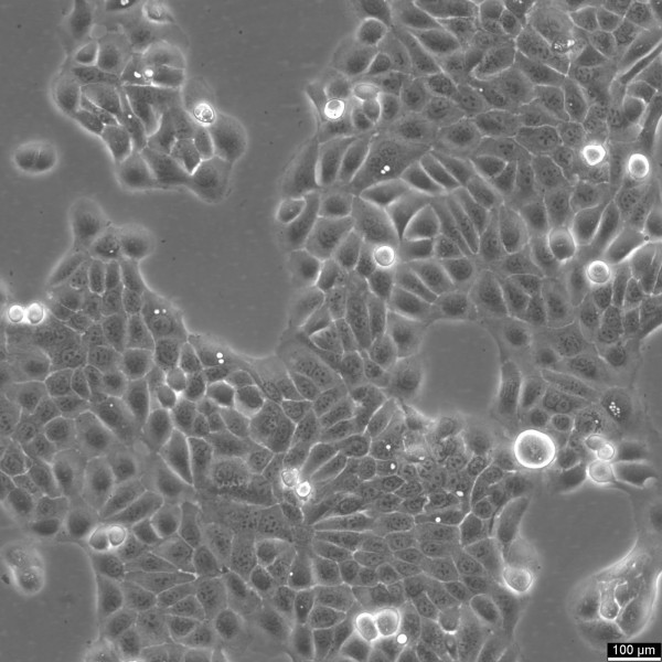
Categories: HaCaT cell lines Description: HaCaT cells are a pivotal model in dermatological research, offering insights into the complex mechanisms of skin biology and pathology. The spontaneously immortalized HaCaT cell line is derived from adult human epidermal cells and retains the capacity to proliferate and undergo differentiation, similar to basal keratinocytes in vivo. HaCaT cells serve as a robust platform for investigating the epidermal differentiation process and studying the epidermal differentiation markers essential for maintaining skin integrity. The susceptibility of HaCaT cells to apoptosis and their sensitivity to apoptosis-inducing agents are extensively studied, particularly in the context of cytotoxic agents like RIPL. Researchers assess these agents' cytotoxicities and the extent of cytotoxicity using HaCaT cells, utilizing techniques such as fluorescence microscopy to visualize cellular changes. Researchers have leveraged HaCaT cells to examine the effects of various agents, including antimicrobial substrates and their influence on cell viability. These cells are an excellent substrate for testing antimicrobial biomaterials and antimicrobial atelocollagen substrates, crucial for skin repair and medical applications. The HaCaT epidermal line also plays a crucial role in studying cellular senescence, cytokines, and gene expression profiles related to aging and chronic diseases. The transcriptional profiles of HaCaT cells, including the role of kappaB and microRNAs, provide insight into the regulatory mechanisms at the molecular level. The HaCaT keratinocyte line, with their characteristics as epidermal keratinocytes, offers a tractable system for dissecting the intricate interplay between epidermal cells and the immune system, specifically the role of keratinocytes in disease states. They enable the exploration of epigenetic modifications and their influence on the differentiation of keratinocytes, including the formation of the cornified envelope, a key feature in the skin's barrier function. In summary, HaCaT cells are an indispensable model in dermatological research, facilitating a deeper understanding of skin biology and pathology through their resemblance to basal keratinocytes and their ability to undergo cell growth and differentiation. Their application spans from studying epidermal differentiation and antimicrobial effects to exploring cellular responses such as apoptosis, making them a cornerstone in cell biology and biomedical research. Organism: Human Tissue: Skin Age: 62 years Gender: Male Ethnicity: Caucasian Cell Type: Keratinocytes with a diameter of 20-25 micrometer. Growth Properties: Adherent Citation: HaCaT (Cytion catalog number 300493) Biosafety Level: 1 Ncbi_ Taxid: 9606.0 Cellosaurus Accession: CVCL_0038 Tumorigenic: No Karyotype: Aneuploid (hypotetraploid) Culture Medium: DMEM, w: 4.5 g/L Glucose, w: 4 mM L-Glutamine, w: 3.7 g/L NaHCO3, w: 1.0 mM Sodium pyruvate (Cytion article number 820300a) Supplements: Supplement the medium with 10% FBS Dissociation Reagent: The 1:1 mixture of EDTA (stock. 0.05%) and trypsin (stock: 0.1%) must be prepared each time ahead of detaching the cells using PBS without Ca2+ and Mg2+ to provide a physiologic osmolarity. Ready-to-use mixtures of trypsin/EDTA are not recommended, as this may result in cell clumps. As an alternative, TrypLE Express (Life Technologies) instead of trypsin/EDTA can be used. The protocol of the manufacturer should be followed. Doubling Time: The doubling time of HaCaT cells is 28 hours. Subculturing: Discard Old Medium: Carefully remove the old culture medium from the flasks. Wash Cells: Add 3-5 ml of phosphate-buffered saline (PBS) without calcium and magnesium to T25 flasks, or 5-10 ml to T75 flasks, to rinse the adherent cells. Add EDTA Solution: Cover the cell layer entirely with a freshly prepared 0.05% EDTA solution. Use 1-2 ml for T25 flasks and 2.5 ml for T75 flasks. Incubate: Incubate the flasks at 37°C for 10 minutes. Add Trypsin/EDTA or TrypLE Express Solution: After incubation, add a freshly prepared trypsin/EDTA solution (0.05% trypsin, 0.025% EDTA) or TrypLE Express to the flasks, ensuring the cell layer is fully covered. Use 1 ml for T25 flasks and 2.5 ml for T75 flasks. (Note: Steps 3 and 4 can be omitted if using TrypLE Express.) Monitor Detachment: Observe the cells under a microscope. The cells should detach within 1-5 minutes. Neutralize Trypsin: Add cell culture medium containing fetal bovine serum (FBS) to neutralize the trypsin activity as soon as the cells have detached. Transfer Cells: Dispense the cell suspension into new flasks pre-filled with fresh culture medium. Seeding Density: 1 x 10⁴ cells/cm2 Fluid Renewal: 2 times per week Freeze Medium: As a cryopreservation medium, use complete growth medium (including FBS) + 10% DMSO for adequate post-thaw viability, or CM-1 (Cytion catalog number 800100), which includes optimized osmoprotectants and metabolic stabilizers to enhance recovery and reduce cryo-induced stress. Thawing And Culturing Cells: Confirm that the vial remains deeply frozen upon delivery, as cells are shipped on dry ice to maintain optimal temperatures during transit. Upon receipt, either store the cryovial immediately at temperatures below -150°C to ensure the preservation of cellular integrity, or proceed to step 3 if immediate culturing is required. For immediate culturing, swiftly thaw the vial by immersing it in a 37°C water bath with clean water and an antimicrobial agent, agitating gently for 40-60 seconds until a small ice clump remains. Perform all subsequent steps under sterile conditions in a flow hood, disinfecting the cryovial with 70% ethanol before opening. Carefully open the disinfected vial and transfer the cell suspension into a 15 ml centrifuge tube containing 8 ml of room-temperature culture medium, mixing gently. Centrifuge the mixture at 300 x g for 3 minutes to separate the cells and carefully discard the supernatant containing residual freezing medium. Gently resuspend the cell pellet in 10 ml of fresh culture medium. For adherent cells, divide the suspension between two T25 culture flasks, for suspension cultures, transfer all the medium into one T25 flask to promote effective cell interaction and growth. Adhere to established subculture protocols for continued growth and maintenance of the cell line, ensuring reliable experimental outcomes. Sterility: Mycoplasma contamination is excluded using both PCR-based assays and luminescence-based mycoplasma detection methods. To ensure there is no bacterial, fungal, or yeast contamination, cell cultures are subjected to daily visual inspections. Safety Precautions: When planning to store a cryovial in liquid nitrogen for future thawing, it is mandatory to adhere to stringent safety measures. Appropriate protective gloves and clothing are essential, and the use of a face mask or safety goggles is required during the transfer of frozen samples to or from the liquid nitrogen tank. This is to mitigate the risk of injury from potential cryovial explosions upon removal, which can result in the projection of sharp fragments. Warranty: We stand by the promise of delivering products with high cell viability and robust culture performance. To achieve the best results, please make sure you follow the storage and culture instructions detailed in the product information sheet closely. Your adherence to these guidelines is key to success. Subject To Third- Party Agreements: Please note that this cell line is not available under a standard Cytion MTA, as it requires a third-party agreement and/or is subject to negotiation with the original licensor. Required Product 1: 820300a Required Product 3: 860015.0 Required Product 4: 830100.0
| Supplier: | Cytion |
| Supplier-Nr: | 300493 |
Properties
| Host: | Human |
| Species reactivity: | human |
Database Information
Handling & Safety
| Storage: | Liquid nitrogen |
| Shipping: | -80°C (International: -80°C) |
Caution
Our products are for laboratory research use only: Not for administration to humans!
Our products are for laboratory research use only: Not for administration to humans!
You will get a certificate here
Viewed