
Cookie-Einstellungen
Diese Website benutzt Cookies, die für den technischen Betrieb der Website erforderlich sind und stets gesetzt werden. Andere Cookies, die den Komfort bei Benutzung dieser Website erhöhen, der Direktwerbung dienen oder die Interaktion mit anderen Websites und sozialen Netzwerken vereinfachen sollen, werden nur mit Ihrer Zustimmung gesetzt.
Konfiguration
Technisch erforderlich
Diese Cookies sind für die Grundfunktionen des Shops notwendig.
"Alle Cookies ablehnen" Cookie
"Alle Cookies annehmen" Cookie
Ausgewählter Shop
CSRF-Token
Cookie-Einstellungen
FACT-Finder Tracking
Individuelle Preise
Kundenspezifisches Caching
Session
Währungswechsel
Komfortfunktionen
Diese Cookies werden genutzt um das Einkaufserlebnis noch ansprechender zu gestalten, beispielsweise für die Wiedererkennung des Besuchers.
Facebook-Seite in der rechten Blog - Sidebar anzeigen
Merkzettel
Statistik & Tracking
Endgeräteerkennung
Kauf- und Surfverhalten mit Google Tag Manager
Partnerprogramm

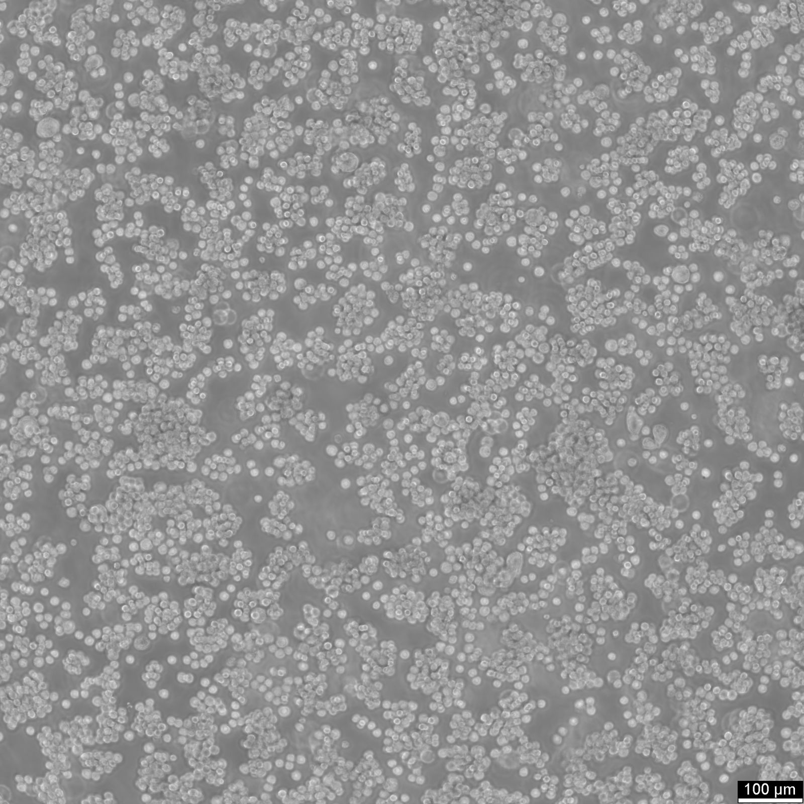
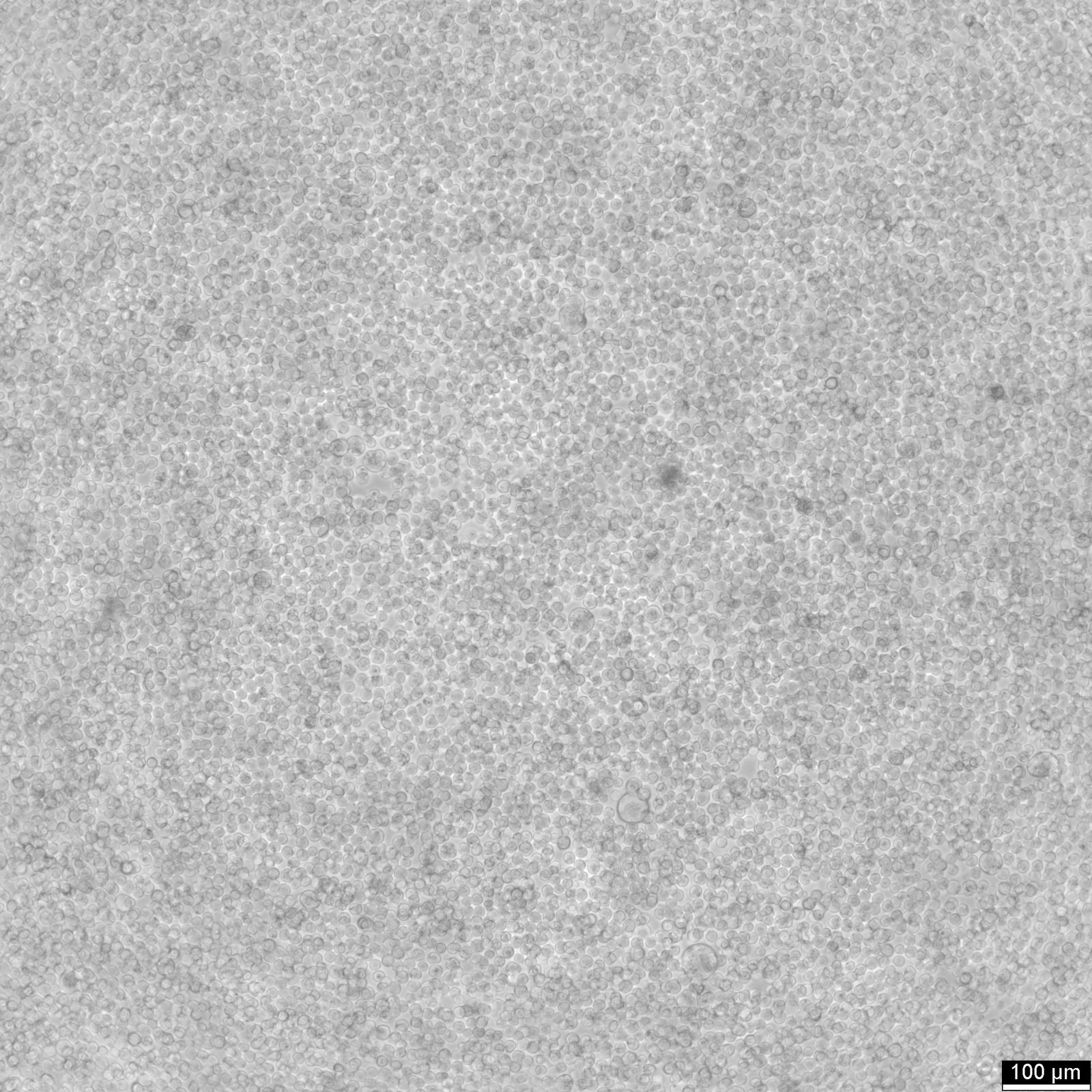
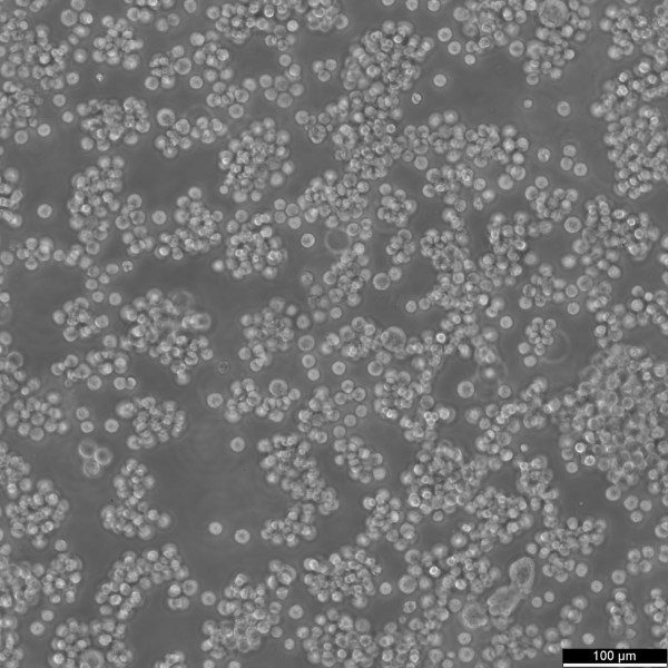


Bitte füllen Sie das Material Transfer Agreement (MTA) aus und reichen Sie es zusammen mit Ihrer Bestellung ein. Für alle kommerziellen Anwendungen füllen Sie bitte das Formular zur beabsichtigten Verwendung aus.
| Artikelnummer | Größe | Datenblatt | Manual | SDB | Lieferzeit | Menge | Preis |
|---|---|---|---|---|---|---|---|
| CYT-305510 | 1 each | - |
3 - 8 Werktage* |
550,00 €
|
Bei Fragen nutzen Sie gerne unser Kontaktformular.
Bestellen Sie auch per E-Mail: info@biomol.com
Größere Menge gewünscht? Bulk-Anfrage
Bestellen Sie auch per E-Mail: info@biomol.com
Größere Menge gewünscht? Bulk-Anfrage
Categories: Lymphoma cell lines Description: The AKATA cell line, derived from Burkitt's... mehr
Produktinformationen "AKATA Cells"
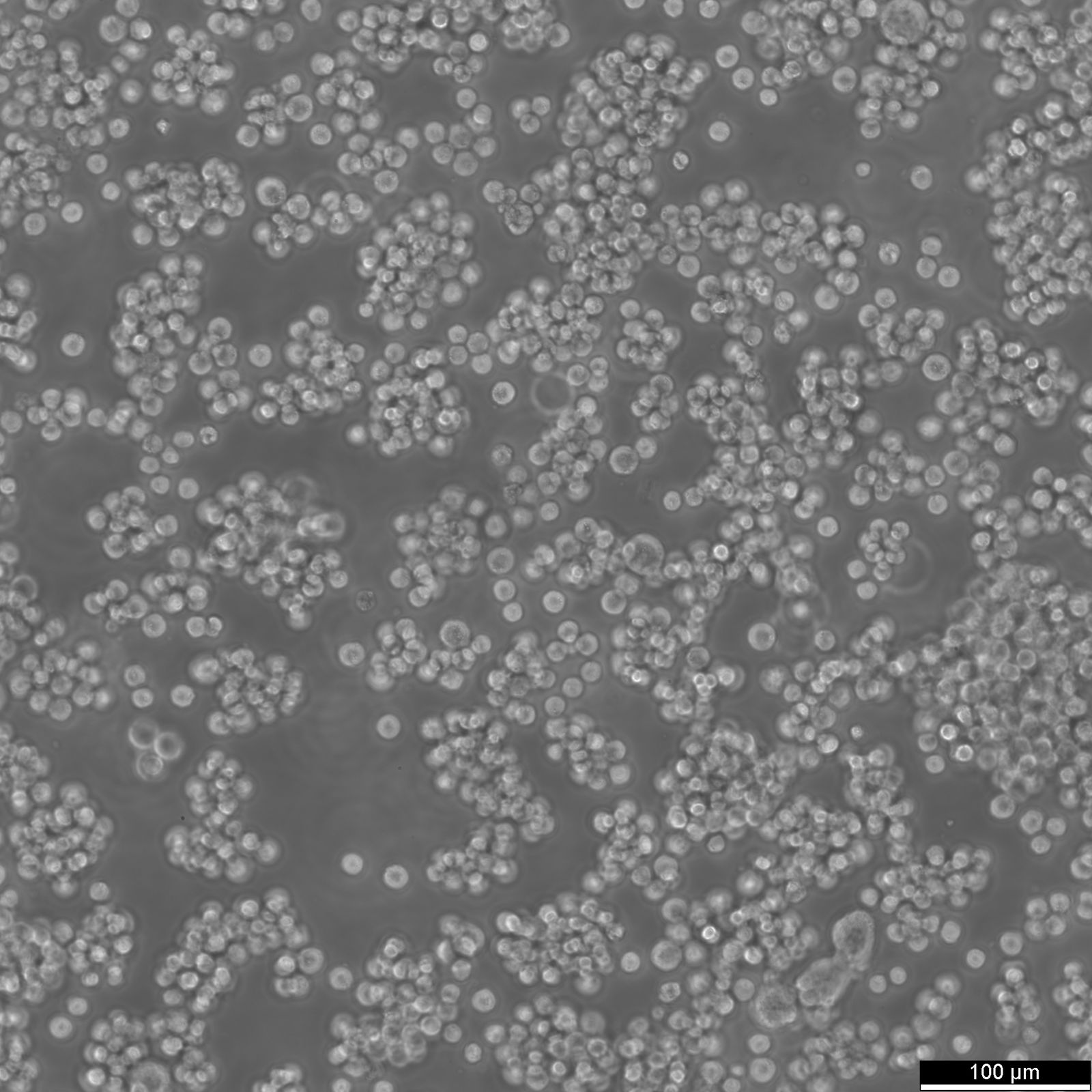
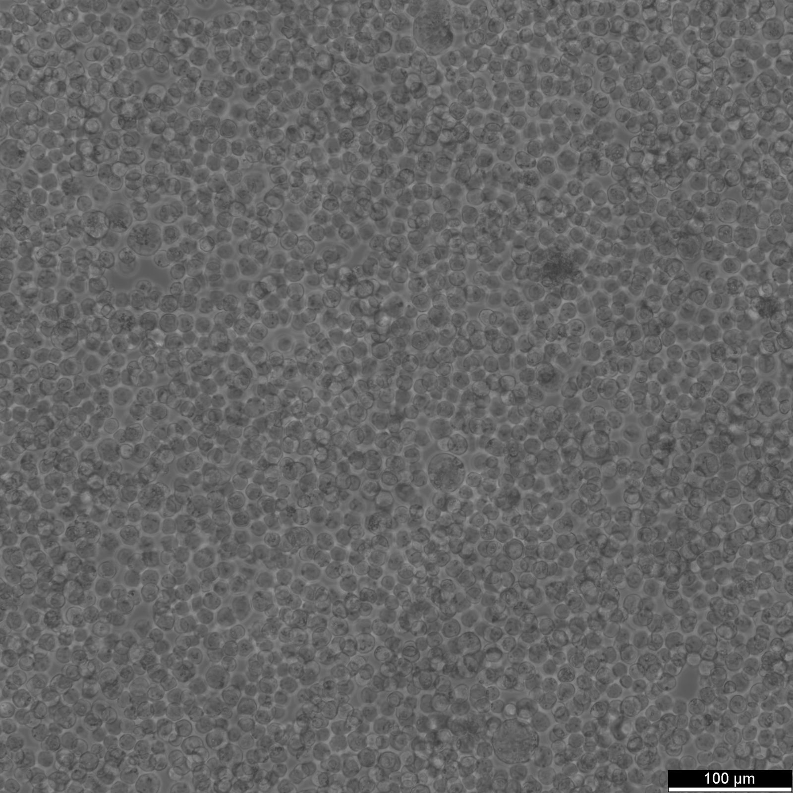
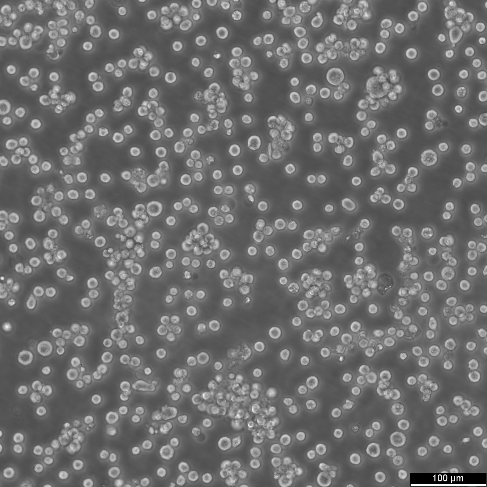
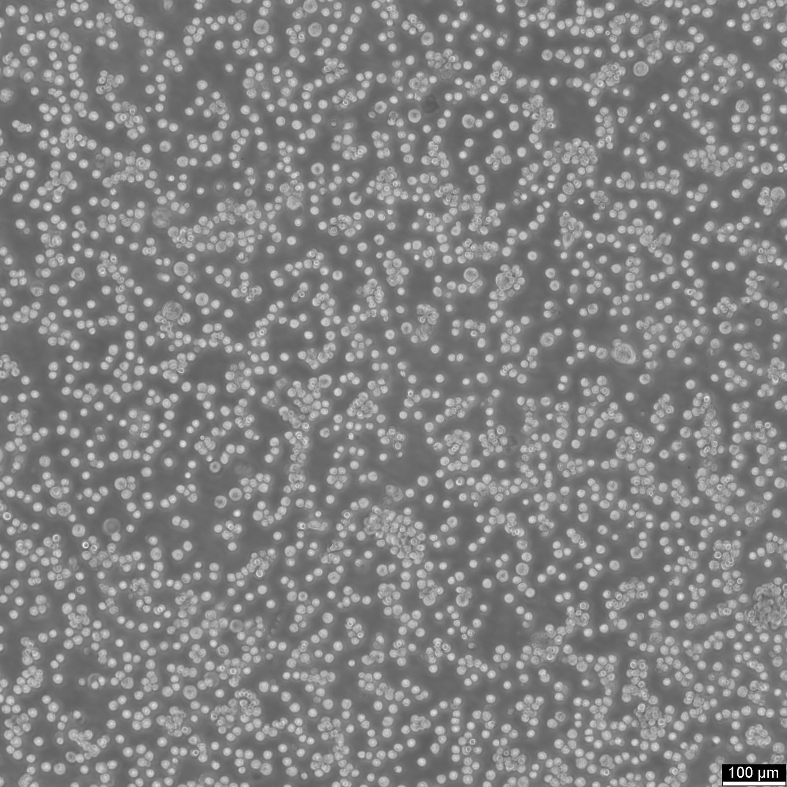
Categories: Lymphoma cell lines Description: The AKATA cell line, derived from Burkitt's lymphoma, is a widely used model to study Epstein-Barr virus (EBV) latency and reactivation. EBV is a ubiquitous herpesvirus linked to a range of cancers, including Burkitt lymphoma, and typically establishes a latent infection within B cells. In AKATA cells, EBV is maintained in an episomal state with a type I latency program, expressing a limited set of viral genes such as EBNA-1, EBER RNAs, and BamHI-A rightward transcripts (BARTs). This restricted gene expression allows the virus to persist in the host without initiating a full lytic cycle. However, AKATA cells can be triggered to enter the lytic phase, where the virus actively replicates and produces progeny. This reactivation is commonly induced through cross-linking surface immunoglobulins, which makes AKATA cells an excellent tool for studying EBV reactivation dynamics and viral gene regulation. Research utilizing the AKATA cell line has also examined the impact of chemotherapeutic agents on EBV reactivation. For instance, drugs like etoposide and doxorubicin have been shown to influence viral latency. Etoposide induces apoptosis in AKATA cells but reactivates EBV less effectively than doxorubicin, which promotes higher levels of lytic gene expression and viral progeny production. Additionally, studies involving gene editing techniques, such as CRISPR/Cas9, have explored the role of epigenetic regulators in AKATA cells. For example, knockout of the histone methyltransferase EZH2 in AKATA cells disrupts the maintenance of latency by reducing the trimethylation of histone H3K27, leading to increased expression of both latent and lytic EBV genes, as well as enhanced viral replication and cell proliferation. AKATA cells also display distinct phenotypic characteristics based on EBV presence, such as increased sensitivity to apoptosis-inducing agents and variations in gene expression related to apoptotic pathways. These differences make EBV-positive AKATA cells a powerful model for dissecting EBV's influence on host cell survival, gene expression, and the virus's lifecycle, particularly in the context of cancer development and potential therapeutic interventions targeting EBV-associated malignancies. Organism: Human Tissue: Blood Disease: Burkitt lymphoma Synonyms: Akata, Akata-BL, Akata BL, Akata-EC, Akata-Early Culture Age: 4 years Gender: Female Ethnicity: Japanese Morphology: Lymphoblast Cell Type: B cell Growth Properties: Suspension Citation: AKATA (Cytion catalog number 305510) Biosafety Level: 2 Ncbi_ Taxid: 9606.0 Cellosaurus Accession: CVCL_0148 Viruses: Transformant: EBV Culture Medium: RPMI 1640, w: 2.0 mM stable Glutamine, w: 2.0 g/L NaHCO3 (Cytion article number 820700a) Supplements: Supplement the medium with 10% FBS Subculturing: Gather the suspension cells in a 15 ml tube and gently wash the adherent cells with PBS lacking calcium and magnesium (use 3-5 ml for T25 flasks and 5-10 ml for T75 flasks). Apply Accutase (1-2 ml for T25 flasks, 2.5 ml for T75 flasks) ensuring full coverage of the cell layer. Allow the cells to incubate at room temperature for 10 minutes. Following incubation, combine and centrifuge both the suspension and adherent cells. After centrifugation, carefully resuspend the cell pellet and transfer the cell suspension into new flasks containing fresh medium. Freeze Medium: As a cryopreservation medium, use complete growth medium (including FBS) + 10% DMSO for adequate post-thaw viability, or CM-1 (Cytion catalog number 800100), which includes optimized osmoprotectants and metabolic stabilizers to enhance recovery and reduce cryo-induced stress. Thawing And Culturing Cells: Confirm that the vial remains deeply frozen upon delivery, as cells are shipped on dry ice to maintain optimal temperatures during transit. Upon receipt, either store the cryovial immediately at temperatures below -150°C to ensure the preservation of cellular integrity, or proceed to step 3 if immediate culturing is required. For immediate culturing, swiftly thaw the vial by immersing it in a 37°C water bath with clean water and an antimicrobial agent, agitating gently for 40-60 seconds until a small ice clump remains. Perform all subsequent steps under sterile conditions in a flow hood, disinfecting the cryovial with 70% ethanol before opening. Carefully open the disinfected vial and transfer the cell suspension into a 15 ml centrifuge tube containing 8 ml of room-temperature culture medium, mixing gently. Centrifuge the mixture at 300 x g for 3 minutes to separate the cells and carefully discard the supernatant containing residual freezing medium. Gently resuspend the cell pellet in 10 ml of fresh culture medium. For adherent cells, divide the suspension between two T25 culture flasks, for suspension cultures, transfer all the medium into one T25 flask to promote effective cell interaction and growth. Adhere to established subculture protocols for continued growth and maintenance of the cell line, ensuring reliable experimental outcomes. Sterility: Mycoplasma contamination is excluded using both PCR-based assays and luminescence-based mycoplasma detection methods. To ensure there is no bacterial, fungal, or yeast contamination, cell cultures are subjected to daily visual inspections. Safety Precautions: When planning to store a cryovial in liquid nitrogen for future thawing, it is mandatory to adhere to stringent safety measures. Appropriate protective gloves and clothing are essential, and the use of a face mask or safety goggles is required during the transfer of frozen samples to or from the liquid nitrogen tank. This is to mitigate the risk of injury from potential cryovial explosions upon removal, which can result in the projection of sharp fragments. Warranty: We stand by the promise of delivering products with high cell viability and robust culture performance. To achieve the best results, please make sure you follow the storage and culture instructions detailed in the product information sheet closely. Your adherence to these guidelines is key to success. Subject To Material Transfer Agreements: If you intend to use Cytion cell lines solely for internal research at a single research site, please complete and sign our Material Transfer Agreement (MTA) and submit it along with your order.For any commercial applications - including but not limited to fee-for-service work, quality control testing, product release, diagnostic use, or regulatory studies - please complete the Intended Use Form so we can prepare a suitable agreement tailored to your project.Please note: The MTA applies only to certain cell lines. If this notice and the MTA document appear on a product page, the agreement is applicable. For cell lines not covered by the MTA, no reference to the agreement will be shown. The MTA is not valid for customers in the Americas, China, or Taiwan. Please contact our U.S. entity to receive the appropriate agreement. Required Product 1: 820700a Required Product 3: 860015.0 Required Product 4: 830100.0
| Hersteller: | Cytion |
| Hersteller-Nr: | 305510 |
Eigenschaften
| Wirt: | Human |
| Spezies-Reaktivität: | human |
Datenbank Information
Handhabung & Sicherheit
| Lagerung: | Liquid nitrogen |
| Versand: | -80°C (International: -80°C) |
Achtung
Nur für Forschungszwecke und Laboruntersuchungen: Nicht für die Anwendung im oder am Menschen!
Nur für Forschungszwecke und Laboruntersuchungen: Nicht für die Anwendung im oder am Menschen!
Hier kriegen Sie ein Zertifikat
Loggen Sie sich ein oder registrieren Sie sich, um Analysenzertifikate anzufordern.
Bewertungen lesen, schreiben und diskutieren... mehr
Kundenbewertungen für "AKATA Cells"
Bewertung schreiben
Loggen Sie sich ein oder registrieren Sie sich, um eine Produktbewertung abzugeben.
Zuletzt angesehen